Night shifts, high volumes, and missed fractures on X-rays

- Pauline Beaumel, AZmed

Radiology departments are under more pressure than they have ever been. Imaging volumes continue to grow year over year while the radiologist workforce struggles to keep pace, a gap that will widen further through 2030 and is identified as a core driver of diagnostic risk in emergency settings.⁶
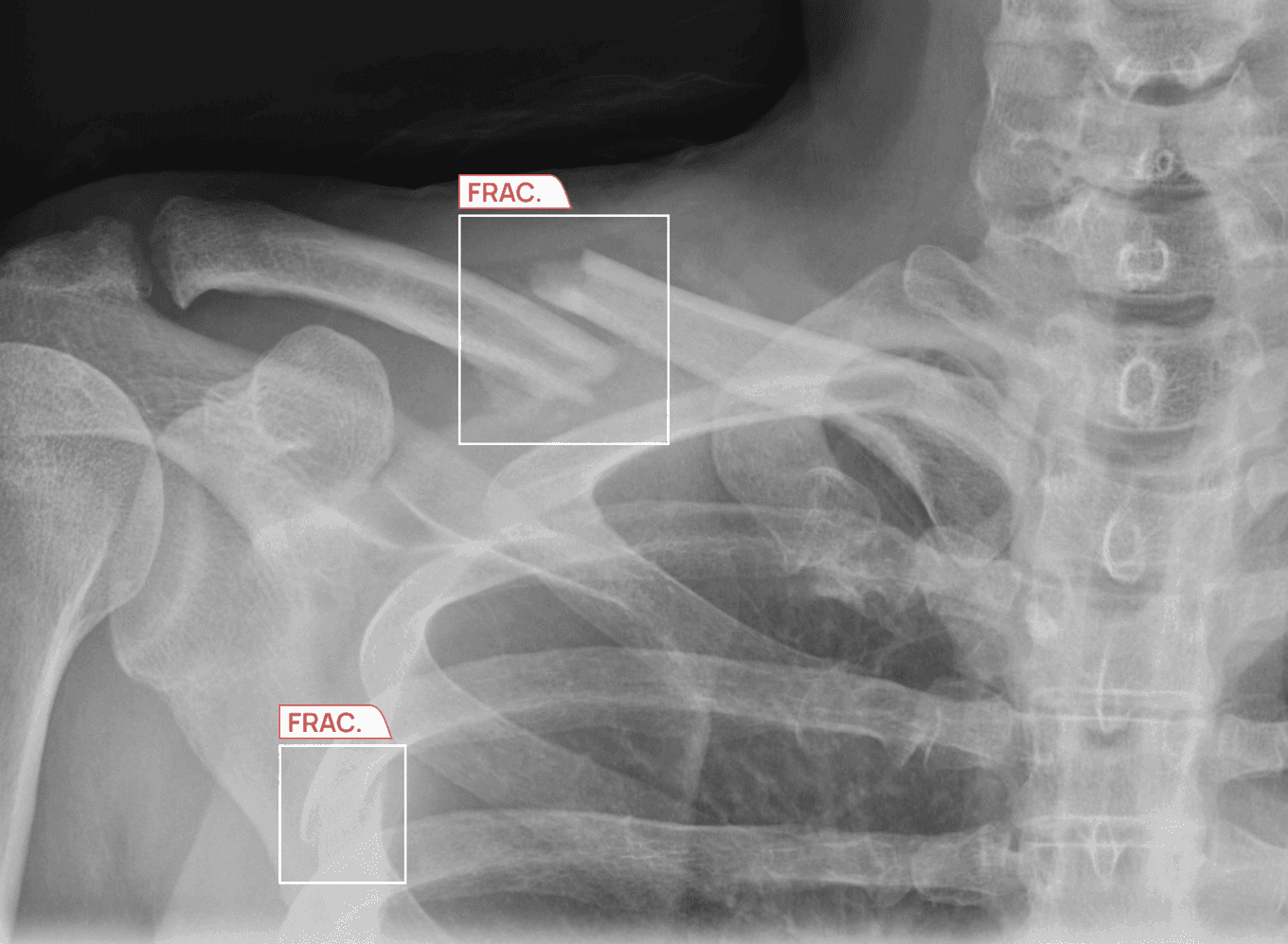
Missed fractures on X-rays sit at the intersection of these pressures, a clinical problem that is well-documented, measurably costly, but increasingly addressable.¹ ²
This article examines why missed fractures happen, when they are most likely to occur, and how AI detection tools are beginning to close the gap without displacing the radiologists who remain crucial to the diagnostic process.
Key takeaways
- Missed fractures on X-rays are usually due to high volume, limited time per case, and overnight coverage gaps.
- Fatigue changes how radiologists search images, increasing review time and the risk of overlooking subtle findings.⁵
- AI supports radiologists as a second reader, helping prioritize urgent cases and improving the detection of subtle fractures without adding workflow friction.¹ ⁷
- AZmed’s Rayvolve® AI Suite, including AZtrauma, provides clinically validated support for fracture detection, helping departments reduce turnaround time while maintaining diagnostic accuracy.¹⁰ ¹³
When radiologists miss fractures
Consider a senior radiologist working an overnight shift at a busy emergency department. The case in front of them is a standard foot X-ray on a 54-year-old who rolled her ankle. It is a routine X-ray, and the radiologist considers a probable soft-tissue injury, clears it, and moves on as the overnight worklist continues to build.
Three days later, the patient returns with significant pain.
A repeat X-ray, this time read during the day, confirms a non-displaced fracture of the fifth metatarsal base, exactly the kind of subtle cortical break that is easy to overlook, especially when attention has been stretched across dozens of cases and the radiologist is operating at reduced efficiency.
Cases like these are quite common.
In most instances, missed fractures are due to a structural failure at the department level, not an individual one. But to understand the scale of this issue, let’s look at some data.
The scale of missed fractures on X-rays in emergency radiology
Studies confirm that the fracture miss rate in the emergency department ranges from 3% to 10%. On its own, this figure may sound modest. But when you apply this percentage to a regional hospital or a large healthcare center that handles thousands of X-rays daily, it results in a substantial number of missed fractures on X-rays, which also has a direct impact on patient care and safety.¹
Patients may return with severe pain or worsened conditions, which can lead to a long-term impact on their health.
In fact, 75% of the malpractice claims against radiologists relate to diagnostic X-ray interpretation error. So, missed fractures on X-rays also increase the time and resources hospitals spend managing these lawsuits.²
Why are foot, elbow, and wrist fractures disproportionately affected?
Some of the most frequent cases of missed fractures involve the foot, elbow, and wrist. This is because the anatomy of these regions is quite complex and genuinely challenging to interpret on plain film. Here’s why:³
- The foot and ankle are composed of more than 26 bones arranged in overlapping projections.
- The elbow presents complex ossification centers, especially in pediatric patients, that can mimic fracture lines.
- The wrist contains the scaphoid bone, whose fractures are usually occult, or hidden, in the acute phase. However, the consequences of missing this type of fracture often lead to avascular necrosis, which is among the most serious complications in orthopedic trauma.
Another factor that increases the fracture miss rate in an emergency department is satisfaction-of-search error. It is a cognitive tendency to stop analyzing an image once a plausible finding has been identified, potentially overlooking secondary lesions on the same film.
So, if a radiologist finds a distal radius fracture, they may stop analyzing and miss a subtle scaphoid fracture in the same wrist study.
Night shifts and missed fractures on X-rays: What the evidence shows
Studies confirm that the risk of missed fractures on X-rays is not evenly distributed across the 24-hour clock. Research published in BMC Emergency Medicine found that 47% of missed fracture cases occurred during the overnight window of 8 pm to 2 am, compared with approximately 20% of correctly diagnosed cases in the control group.⁴
That concentration of errors in a 6-hour window is due to specific factors.
- Radiologist coverage is reduced or entirely absent at many centers, with overnight reading handed to on-call radiologists working in isolation.
- Junior clinicians manage more cases with less immediate senior support.
- Worklists that have accumulated since the end of the afternoon shift arrive in sequence, regardless of clinical urgency, creating a first-in, first-out queue that places routine cases alongside time-sensitive trauma cases.
The disruption of the human circadian rhythm during the night also increases the risk of errors and missed diagnoses. Let’s understand this in more detail below.
How fatigue changes visual search patterns for radiologists
Radiologist fatigue during the overnight shift is known to increase cases of missed fractures. A study available through the National Library of Medicine quantifies this.⁵
Research using eye-tracking technology found that radiologists working overnight shifts made 60% more gaze fixations than radiologists working during the day when reviewing the same images.⁵
It took the night-shift radiologists approximately 34% longer to locate a fracture when one was present.⁵
The study associated these outcomes with 5 fatigue-related factors: lack of energy, physical exertion, physical discomfort, lack of motivation, and sleepiness. These symptoms can contribute to a decrease in emergency radiology diagnostic accuracy.
But this is just one part of the problem. Another factor that leads to missed fractures on X-rays is the ever-increasing number of cases that radiologists have to deal with every day.
High imaging volumes compound missed fracture risk
Radiologists must read an increasing number of X-ray images every day. For example, in 2023, SimonMed, which operates over 150 outpatient imaging centers across the United States, reported that the volume of trauma X-rays increased by 20% compared with 2022 figures, with approximately 100,000 additional trauma X-rays being read in a single year.¹⁰
This increase in volume has continued in 2026, and the U.S. has a shortage of radiologists, meaning each radiologist must read more X-rays every day and has less time per X-ray than ever before. The EU-REST project, an effort funded by the European Commission to collect data on staffing, training, and equipment in Europe, states that this shortage could lead to delays in providing emergency treatment.⁶
The gap between the number of X-rays and radiologists is expected to grow significantly by 2030; therefore, the number of missed fractures on X-rays could grow significantly as well.⁶
As a result, many hospitals are looking at different options to help reduce radiologist stress and missed fractures, including AI-assisted tools.
How AI reduces missed fractures without replacing the radiologist
AI tools in radiology analyze X-rays for potential fractures by acting as a second set of eyes. They highlight areas on the X-ray that may need attention, specifically improving sensitivity to subtle fractures such as avulsion or buckle fractures. This is possible because of advances in deep learning and automated diagnostics in musculoskeletal radiology.¹ ⁸ ⁹
Although AI can help reduce missed fractures, the purpose of using digital image processing software in radiography is not to remove the radiologist from the process, but to enhance their efficiency and workflow, especially in busy emergency radiology settings, through worklist prioritization and triage to minimize the chance of human error.⁷ ⁹
How it works in practice:
- The AI system searches through all the images it receives for features indicative of fracture, such as cortical discontinuities, trabecular disruptions, and soft-tissue swelling.
- If the AI system identifies these features with a level of confidence above a set threshold, it flags the case for review by the radiologist.
- The radiologist reviews the flagged findings, determines whether the fracture is present, and completes the report.
Worklist triaging: Prioritizing critical cases first
Normally, when radiologists interpret X-rays, they do so on a first-in, first-out basis. Therefore, on any given day, due to large volumes of X-ray requests compared with available radiologists to interpret them, a routine X-ray may be interpreted before an X-ray with a critical finding. This is especially true late in the day when there is no senior reviewer to assign critical cases for review.
AI assists with this because it flags cases that require immediate review above routine requests.⁷
For more on X-ray worklists, please review our complete guide.
AI functions as a trustworthy second reader on every shift
As discussed earlier, overnight fatigue can reduce diagnostic performance. AI plays a significant role in this context because it does not experience the same visual and cognitive fatigue that occurs for people after performing the same task for 8 hours consecutively.
A prospective study offers relevant evidence. An independent side-by-side comparison of 3 commercial AI solutions was performed by TUM University Hospital Rechts der Isar in Munich, Germany, and compared the tools with radiologist reports. The study was published in Radiography in October 2025.⁸
In that Radiography article, AZmed's AI-powered platform had the highest accuracy score, with an AUC of 84.88%, and the highest sensitivity score, at 79.48%, among the AI systems for detecting fractures.⁸
AZmed's high level of sensitivity gives it value as a first-line screening tool.⁸
This is important in a high-volume radiology workflow setting where a missed diagnosis could have significant clinical and operational implications.
However, although AI tools showed relatively lower accuracy in complex cases, the tools aid radiologists by reducing their workload, serving as a second reader, and flagging urgent cases for radiologists to review.⁸ ⁹
Evidence from clinical practice before and after AI implementation
AI-powered radiology products, including those under the Rayvolve® AI Suite, have been implemented in over 2,500 healthcare centers around the world. Hospitals that implement AI detection have experienced substantial improvements in TAT, which reflects how long it takes from image capture until the final report is delivered.¹⁰ ¹³
As part of a nationwide outpatient imaging network, SimonMed Imaging began using the AZtrauma AI radiology solution to identify fractures, dislocations, and joint effusions on X-rays across its 150 locations.¹⁰
After 1 month
AZtrauma helped radiologists diagnose bone fractures nearly 6 times faster, showing a significant reduction in report turnaround time with AI fracture detection.¹⁰
AZtrauma was also beneficial for distinguishing between routine X-rays and high-priority X-rays, so urgent cases were able to be processed much more quickly than those classified as routine.¹⁰
After 6 months
The tool continued to reduce TAT while maintaining sensitivity and specificity. The percentage of cases diagnosed as acute fractures increased from 10.4% without AI to 11.8% with AI.¹⁰
Sean D. Raj, CIO at SimonMed Imaging, stated:
“Based on our study, since the implementation of the AI, a patient diagnosed with a fracture receives results 6 times faster.”¹⁰
View the full use of AI in fracture detection here.
What to evaluate when using AI for missed fracture detection
Radiology departments and hospital administrative leaders evaluating AI fracture detection tools should begin with clinical evidence, but it should not be the only criterion.¹¹
Here are several practical recommendations to help you select the top AI for fracture detection.
- External validation: Is there sufficient research showing that the AI tool can perform well beyond pilot stages? For example, tools under the Rayvolve® AI Suite have multiple scientific studies that demonstrate effectiveness in day-to-day applications and real-world settings. Peer-reviewed medical literature should be reviewed before making a purchasing decision. Information supplied only by an AI vendor is not sufficient.¹¹ ¹³
- Pediatric coverage: When reading pediatric X-ray films, it is more difficult to correctly assess whether fractures are present because a child's bones are still developing and can mimic fracture lines in some cases. Because of this, you want to find an imaging tool that takes into account the biological differences of children.¹¹
- PACS integration: If a radiology tool does not reduce radiologists' workload, then that tool is not beneficial. The best radiology tools are those that integrate with the PACS in your facility and are used within the radiologist’s existing workflow.¹¹
- Regulatory clearance: When you are reviewing tools in terms of FDA or CE regulatory approvals for clinical use, you must verify vendor documentation, including copies of regulatory clearance or certification and any additional information that may be available from the vendor.¹²
Missed fractures on X-rays as a solvable problem
AI fracture detection tools like AZtrauma are making it possible to better identify and manage fractures, shorten turnaround times for urgent cases, and provide a consistent level of diagnostic support across shifts that no staffing model can replicate through human coverage alone.¹⁰ ¹³
The radiologist remains the clinical decision-maker. With this support, radiologists can make patient-care decisions with the best available information at the time.
For additional clinical documentation to validate AZtrauma as an AI-based tool for fracture detection, please visit azmed.co/resources/scientific-evidence.¹³
Frequently asked questions
What is the typical fracture miss rate in emergency departments?
Fracture miss rates for emergency department X-ray exams range from 3% to 10%, depending on the number of cases and overall reporting conditions.¹
Why are fractures commonly missed on X-rays?
Missed fractures on X-ray examinations can occur due to subtle variations in presentation, increased complexity of the anatomy, time pressure on the radiologist, and cognitive biases such as satisfaction-of-search error.¹ ³
Does radiologist fatigue affect diagnostic accuracy?
Yes, research indicates that radiologist fatigue negatively affects diagnostic accuracy. Radiologist fatigue can cause slower scanning, increased fixation rates, and reduced sensitivity to identifying minor abnormalities during the night shift.⁵
Are certain anatomical regions more prone to missed fractures?
Yes. Foot, wrist, and elbow fractures are commonly missed on X-rays, primarily due to overlapping bones and fractures that are not readily identifiable.³
What changes in radiologists' visual search during night shifts?
Radiologists' visual search patterns can change while working at night, with eye-tracking studies demonstrating a higher number of fixation events and longer search times compared with other shifts.⁵
How does worklist structure impact missed fractures?
Missed fractures may occur when urgent and routine cases are placed together within a first-in, first-out workflow.⁷
Are junior clinicians more likely to miss fractures overnight?
Most overnight shifts have more limited access to senior staff, which can result in greater variability in detection.⁴
What clinical consequences follow a missed fracture on initial X-ray?
Complications due to a missed fracture can include ongoing pain, delayed treatment, non-union of the fracture, and avascular necrosis.¹
How does AI specifically help during overnight radiology shifts?
By using AI-assisted fracture detection tools, radiologists can detect and prioritize urgent cases more reliably throughout the night, especially when human performance may decline.⁵ ⁷
Can double reading reduce missed fracture rates?
Yes. A second review, whether by another radiologist or AI support, can improve the accuracy of detecting both subtle and multiple fractures.¹ ⁹
References
- Kuo RYL, Harrison C, Curran TA, et al. Artificial Intelligence in Fracture Detection: A Systematic Review and Meta-Analysis. Radiology: Artificial Intelligence. 2022.
- Zhang L, et al. Diagnostic error and bias in the department of radiology. 2023.
- Freed HA, Shields NN. Most frequently overlooked radiographically apparent fractures in a teaching hospital emergency department. Ann Emerg Med. 1984.
- Hallas P, Ellingsen T. Errors in fracture diagnoses in the emergency department: characteristics of patients and diurnal variation. BMC Emergency Medicine. 2006.
- Hanna TN, et al. The Effects of Fatigue from Overnight Shifts on Radiology Search Patterns and Diagnostic Performance. Journal of the American College of Radiology. 2018.
- AZmed. EU-REST project warns that radiologist gap will widen by 2030.
- AZmed. AI Worklist Triage for X-rays Throughput and Case Mix.
- Luiken I, Lemke T, Komenda A, et al. Evaluation of commercial AI algorithms for the detection of fractures, effusions, and dislocations on real-world clinical data: A prospective registry study. Radiography. 2025.
- AZmed. Will AI replace radiologists? What AI radiology can and cannot do.
- AZmed. Impact of AI on Fracture Detection in Radiology.
- AZmed. How to Choose the Best AI for Fracture Detection.
- AZmed. How to evaluate FDA- or CE-cleared AI for routine radiography.
- AZmed. AI Radiology: Clinical Studies & Scientific Evidence.
Regulatory information
US - Medical device Class II according to the 510K clearances. Rayvolve: is a computer-assisted detection and diagnosis (CAD) software device to assist radiologists and emergency physicians in detecting fractures during the review of radiographs of the musculoskeletal system. Rayvolve is indicated for adult and pediatric population (≥ 2 years).Rayvolve PTX/PE: is a radiological computer-assisted triage and notification software that analyzes chest x-ray images of patients 18 years of age or older for the presence of pre-specified suspected critical findings (pleural effusion and/or pneumothorax). Rayvolve LN: is a computer-aided detection software device to assist radiologists to identify and mark regions in relation to suspected pulmonary nodules from 6 to 30mm size of patients of 18 years of age or olderEU - Rayvolve: Medical Device Class IIa in Europe (CE 2797) in compliance with the Medical Device Regulation (2017/745). Rayvolve is a computer-aided diagnosis tool, intended to help radiologists and emergency physicians to detect and localize abnormalities on standard X-rays.Caution: The data mentioned are sourced from internal documents, internal studies and literature reviews. It is for distribution to Health Care Professionals only and should not be relied upon by any other persons. Carefully read the instructions for use before use. Please refer to our Privacy policy on our website For more information, please contact contact@azmed.co.AZmed 10 rue d’Uzès, 75002 Paris - www.azmed.co - RCS Laval B 841 673 601© 2026 AZmed – All rights reserved. MM-26-25