AI radiology supports faster, more accurate diagnosis. Read this blog to discover how radiologists and AI work together and why one isn’t a replacement for the other.
“People should stop training radiologists now. It is just completely obvious that within 5 years, deep learning will do better than radiologists.” That’s what the British computer scientist and Nobel Prize winner, Geoffrey Hinton, said in 2016.
This statement sparked endless debate, with the media quick to paint AI as a replacement rather than a support for medical professionals.
Fast forward to 2025, when the daily reality looks very different. Imaging volumes continue to climb, trauma teams need reads around the clock, and radiology departments everywhere are stretched thin. This pressure shows up in backlogs, longer turnaround times, and the constant risk of fatigue-related errors.
So it’s no surprise that the question lingers: if radiologists can’t keep pace, will machines step in to take their place? The honest answer is no.
What we are seeing instead is a shift toward collaboration: AI working alongside radiologists to make an unmanageable workload manageable, without replacing the expertise that only humans bring to patient care.
Why does the debate persist between AI and radiologists?
Ever since the introduction of AI, the debate about AI replacing a particular profession has been rampant. And this applies to radiology, too.
In 2016, the Nobel Prize winner in Physics, Geoffrey Hinton, stated boldly that AI would completely replace radiologists within 5 years. These claims were broadly correct that the technology would have a significant impact on the job roles of radiologists, but not as a job killer.
The claims of AI replacing radiologists rest on a fundamental misunderstanding of what radiologists actually do.
When performing an imaging exam, radiologists take into account patient history, symptoms, prior imaging, laboratory results, and treatment plans while giving a diagnosis. They also assist medical professionals in other fields through their findings, all the while bearing legal responsibility for their interpretations.
Studies show that there is a growing shortage of radiologists in the US. And that’s where using AI can help healthcare centers.
What can AI do reliably today in radiology?
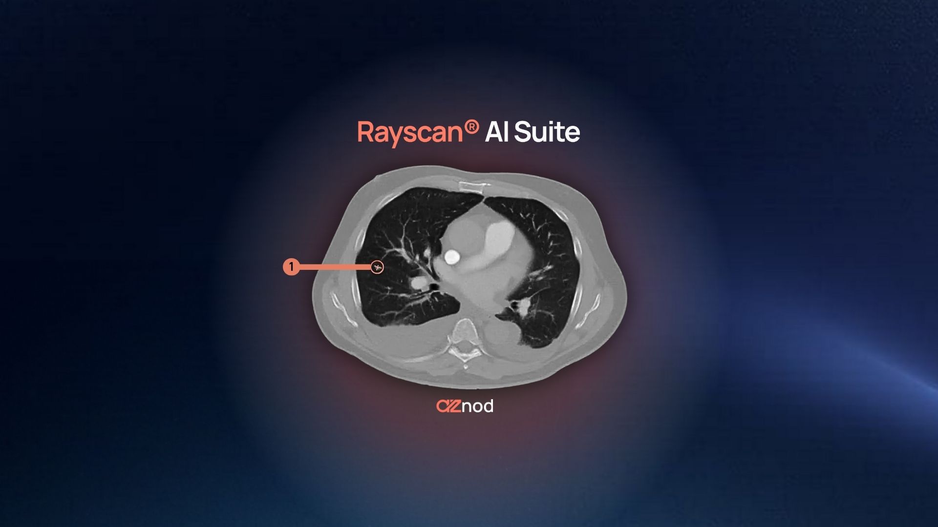
The debate around AI radiology is largely driven by the impressive results of deep learning neural networks in image recognition tasks. AI tools, like those in the Rayvolve® AI Suite, have been able to learn from large amounts of data and have shown remarkable abilities to enable early detection of pathology, reduce false-positive rates that can fatigue the human eye, and guide more precise treatment plans. Here’s how:
- AZtrauma: Part of the Rayvolve® suite, AZtrauma is CE- and FDA- cleared. It is the leading AI radiology solution for detecting fractures, dislocations, and joint effusions on X-rays. AZtrauma is clinically validated by top institutions across the US and helps achieve up to 83% turnaround time reduction (TAT) at healthcare centers.
- * AZchest: This product is CE- and FDA- cleared (for two applications and three indications). It has gained popularity due to its ability to automatically categorize, detect, and report the main cardiac and lung abnormalities on X-rays. It helps radiologists detect pathologies such as pulmonary edema, cardiomegaly, and pleural effusion, among others.
- AZmeasure: AZmeasure delivers automated characterization of osteo-articular geometries, including lengths and angular positions. This CE-marked tool helps in detecting pathologies such as scoliosis, hallux valgus, hip dysplasia and FAI.
- AZboneage: Another CE-marked AI radiology tool that helps radiologists automate the calculation of the bone age of pediatric patients, based on the Greulich & Pyle atlas. It seamlessly integrates into clinical workflows. The product also performs statistical comparisons between the bone and chronological age of patients.
AI radiology tools like these help optimize a lot of the manual tasks that radiologists do throughout their day.
What can AI radiology not do?
Despite the bold statements made in 2016 and the benefits of AI radiology tools, there are a few things only radiologists can do.
Communicate results to patients
A huge part of the job role of any clinicians is to communicate findings to the patients. It is to assure them when they get anxious, discuss next steps, address concerns, and coordinate follow-up. These conversations require empathy, adaptability, and nuanced communication that AI simply can’t provide.
Weigh imaging results against clinical context
Findings in the medical field aren’t simply black or white and often require context before radiologists can suggest the right course of action. While AI can tell that a patient has a lung abnormality on an X-ray, it is the radiologist's job to compare it against prior studies, consider the patient's treatment response, note clinical changes since the last imaging, and adjust their interpretation accordingly.
And if findings seem inconsistent with clinical presentation, radiologists reach out to other professionals to discuss the case.
Take legal responsibility for patient care
Legal and ethical responsibility for patients is yet another sector where AI cannot replace radiologists. Because someone must ultimately stand behind diagnostic decisions, accept accountability for errors, and navigate the medicolegal landscape of medical practice. AI tools simply can’t assume these responsibilities.
Provide nuanced judgment
Similar to the point we discussed above on clinical context, AI also can’t provide nuanced judgment yet. For example, if a patient is diagnosed with pneumothorax and has also suffered from a recent infection, it is the radiologist's experience that would dictate the right course of action. They would prioritize urgent care, suggest next steps, and sometimes acknowledge diagnostic uncertainty rather than forcing conclusions.
This tolerance for ambiguity and ability to reason through incomplete information remains distinctly human because AI is known to hallucinate when it lacks information, which can often lead to wrong diagnoses.
These are all aspects of a radiologist's job that AI can’t replace yet. But as we’ve discussed, AI can definitely reduce manual work so radiologists can focus on these skills rather than generating interpretations.
And that’s why medical centers throughout the world are adopting AI radiology tools. To give back the time radiologists need to deliver better care.
Choosing the right tools can make all the difference.
Clinical validation as the differentiator while choosing AI tools
Ever since the statement made in 2016, there has been a surge of research and development in radiology AI, with hundreds of papers published every year, dozens of start-ups founded, and millions of dollars invested.
But it all comes down to just one question - will the tool work in the day-to-day workflow of a radiologist while giving accurate results?
A tool that works in theory is very different from one that holds up under the pressures of a busy emergency department or the variability of real-world imaging.
That’s why the tools you choose should have regulatory clearance. CE markings and FDA clearances are essential regulatory approvals for medical tools, allowing their sale in the European Union and the United States, respectively. These clearances ensure that the tools have passed rigorous safety and performance standards. But it is one thing that the tool has passed clearance, and it is another that it's being used by radiologists.
AZmed’s Rayvolve® suite (AZtrauma, AZchest, AZmeasure, AZboneage) meets these standards.
These solutions have been clinically validated across continents, and most importantly, are already integrated into the daily workflows of thousands of radiologists. Because they plug directly into PACS/RIS, they don’t add an extra layer of cognitive load or require new systems training. They just work in the background, supporting the way you already practice.
AZmed and radiologists: Building the future together
AI is definitely changing the role of radiologists. But the debate between AI and radiologists isn’t about choosing between humans or machines; it’s about deciding how we can work smarter with the resources we have.
As you’ve seen through AZmed’s tools, AI radiology is well-suited for the repetitive and time-sensitive parts of the job. It can help reduce burnout and make sure that radiologists spend their time doing what they do best - providing patient care.
In an interview with the New York Times, Dr. Halamka, an A.I. optimist, put it best: “Five years from now, it will be malpractice not to use A.I.,” he said. “But it will be humans and A.I. working together.”
And that’s where solutions like Rayvolve® give radiologists the support they need to stay ahead without compromising accuracy or patient trust. That’s the real future of radiology, not replacement, but partnership.
Explore how Rayvolve® helps radiologists to keep pace with rising imaging demand. Book a demo with our team today.
FAQs
- Will AI replace radiologists in the future?
AI will not replace radiologists. AI is a tool that supports radiologists by automating repetitive image analysis, decreasing fatigue, and increasing diagnostic accuracy, but radiologists will always be needed for clinical judgment, patient communication, and the final diagnosis.
- Why do people think AI can replace radiologists?
The idea that AI could replace radiologists arose from the single-minded early projections that deep learning would outperform humans in image interpretation. AI can analyze medical images quickly, but, AI is limited because it does not understand clinical context, it cannot exercise empathy, and it bears no legal accountability.
- What are the limitations of AI in radiology?
AI does not comprehend patient context, deliver care, or assume legal liability for diagnoses. AI also does not work well with ambiguous cases or rare pathologies that require human judgment and compassion, nor can it make ethical judgments about cases or treatment choices.
- How does AI improve radiology workflows?
AI streamlines workflow by triaging critical cases, shortening turnaround times (TAT), and automating measurements and report writing, thereby allowing radiologists to spend time on higher-value activities, such as complex cases and patient care.
- What is the Rayvolve® AI Suite?
Rayvolve® is the CE-marked and FDA-cleared AI suite from AZmed that is designed for use in routine radiography. The Rayvolve® AI Suite comprises four modules: AZtrauma for fracture detection, AZchest for thoracic analysis, AZmeasure for osteoarticular measurements, and AZboneage for bone age estimation in pediatric patients.
- What are CE and FDA clearances in medical AI?
CE marking indicates that medical devices can be marketed and sold in the European Union. FDA clearance means the device is authorized for use in the United States. Both CE marking and FDA clearances are regulatory pathways that indicate the AI tool has met rigorous safety and performance standards, all while undergoing clinical validation.
- How accurate are AI tools in detecting fractures?
In clinical studies, AI algorithms have been shown to obtain sensitivities above 95% in fracture identification. For example, AZtrauma attained 95.6% sensitivity and 91.6% specificity in identifying bone fractures across small, medium, and large data sets.
- What is the role of AI in chest X-ray analysis?
AI tools (e.g., AZchest) automatically detect and classify pulmonary and cardiac abnormalities such as pneumothorax, pleural effusion, and cardiomegaly to facilitate early diagnosis and improve chest X-ray interpretation efficiency.
- Can AI detect subtle or missed findings on X-rays?
Yes. AI can identify subtle pathologies that can sometimes be overlooked during busy shifts. An example is Rayvolve®, which has demonstrated it can identify microfractures (e.g., a stress fracture) and small nodules potentially missed during human interpretation.
- Why is clinical validation important for AI radiology tools?
Clinical validation ensures that AI models generate reliable performance in diverse real-world conditions across diverse patient populations and on different imaging systems. It is possible that the AI tool will not produce consistent or safe diagnostic results without clinical validation.
- What is the difference between AI triage and diagnostic AI?
AI triage places critical cases, such as fractures or pneumothorax, at the top of a radiologist's worklist. Diagnostic AI instead gives structured findings to assist in report preparation and treatment planning.
- How does AI-assisted reporting work in radiology?
AI-assisted reporting automatically pre-fills structured fields, such as “Findings” and “Impressions,” into radiology reports. Radiologists then review and edit the resulting drafts, which saves them from a substantial amount of manual typing while maintaining full clinical authorship of the report.
- How does AI integrate with PACS/RIS systems?
AI solutions such as Rayvolve® integrate directly into existing PACS and RIS systems, which allows for studies requiring triage to be routed automatically to a radiologist, or studies requiring results to be routed automatically to the current careprovider with results automatically visualized; all while limiting the disruption to workflow for the radiologist and/or technologist.
- What legal or ethical limits prevent AI from replacing radiologists?
AI cannot be legally held responsible for any medical diagnosis, nor can it be held responsible for medical risk management relating to the health of a patient. Only licensed physicians can be held responsible for medical decisions, which requires work on behalf of the healthcare provider, with regard to diagnosis and/or methodology for patient prognostication. AI systems are not an adjunct to medical practice intended to substitute for human diagnostic work; AI is intended to be a supplemental tool to a licensed physician.
- Can AI improve diagnostic consistency across radiology departments?
Yes. AI standardizes interpretations by applying the same decision criteria across all cases. This reduces inter-reader variability and ensures consistent reporting across departments, shifts, and sites.
- How widespread is the adoption of AI in radiology today?
AI in radiology is rapidly expanding, with tools like Rayvolve® deployed in more than 2,500 healthcare facilities across over 55 countries. Adoption continues to grow as evidence of safety, accuracy, and workflow benefits increases.
- What is the future of AI and radiology collaboration?
The future lies in collaboration, not replacement. AI will continue to handle repetitive, time-sensitive image analysis, while radiologists focus on clinical reasoning, patient interaction, and decision-making.
Regulatory mentions:
* AZchest is CE-marked for detection of abnormalities on chest X-rays such as: consolidation, pleural effusion, pneumothorax, cardiomegaly, acute pulmonary edema, ribs fractures and pulmonary nodules. It’s also FDA-cleared for two applications: detection of lung nodules (with Rayvolve LN) and notification and triage for pneumothorax and pleural effusion (with Rayvolve PTX-PE)
EU - Rayvolve: Medical Device Class IIa in Europe (CE 2797) in compliance with the Medical Device Regulation (2017/745). Rayvolve is a computer-aided diagnosis tool, intended to help radiologists and emergency physicians to detect and localize abnormalities on standard X-rays.
US - Medical device Class II according to the 510K clearances. Rayvolve: is a computer-assisted detection and diagnosis (CAD) software device to assist radiologists and emergency physicians in detecting fractures during the review of radiographs of the musculoskeletal system. Rayvolve is indicated for the adult and pediatric population (≥ 2 years).
Rayvolve PTX/PE: is a radiological computer-assisted triage and notification software that analyzes chest x-ray images of patients 18 years of age or older for the presence of pre-specified suspected critical findings (pleural effusion and/or pneumothorax). Rayvolve LN: is a computer-aided detection software device to assist radiologists to identify and mark regions in relation to suspected pulmonary nodules from 6 to 30mm size of patients of 18 years of age or older.
Caution: The data mentioned are sourced from internal documents, internal studies and literature reviews. Carefully read the instructions for use before use.
AZmed 10 rue d’Uzès, 75002 Paris - www.azmed.co - RCS Laval B 841 673 601
© 2025 AZmed – All rights reserved. MM-25-37


