Why emergency fractures are most likely to be missed at night

- Pauline Beaumel, AZmed

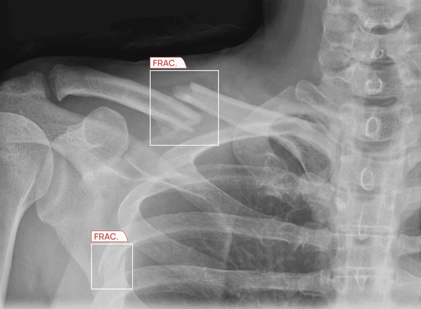
The risk of a missed fracture in the emergency department is highest during evening and overnight hours because patients with very subtle fracture patterns are more likely to present later in the day, when healthcare providers may be fatigued and have limited senior review. This is the problem of missed fractures at night in the emergency department in its simplest form. The purpose of this article is to look at why the pattern of missed fractures at night in the emergency department occurs, using existing clinical studies that suggest that fatigue, low-acuity injuries such as non-displaced or subtle fractures, and staffing patterns contribute to a higher rate of fracture misdiagnosis.
Key takeaways
- Missed fracture diagnoses increase during evening and overnight hours as fatigue builds and senior review decreases. Therefore, after-hours fracture diagnosis should be seen not as an individual error, but as a system-level risk.
- Subtle or low-acuity injuries tend to be presented later in the day, and studies show that missed fracture diagnoses are more common during evening and after-hours periods.
- Fatigue has been shown to alter visual search patterns and lower diagnostic accuracy, even when the radiologist takes longer than usual to complete the case.
- The first interpretation of radiographic images during evening and overnight shifts in most hospitals is performed by less-experienced clinicians, which can reduce diagnostic accuracy.
- Artificial intelligence (AI) tools such as AZtrauma support fracture detection 24/7 by flagging suspected fractures and prioritizing urgent studies for earlier review, including during after-hours periods when staffing and fatigue pressures are higher.
A fracture that waited until morning to be found
One familiar example many emergency departments and hospitals have experienced is this:
A 40-year-old patient presents to your emergency department at 9:30 pm, complaining of wrist pain after a fall. The patient delayed coming in until the evening to complete work because the pain was mild.
The emergency department provider at the time orders an X-ray and, after reviewing the available notes, sees no obvious fracture. The patient is discharged with instructions indicating a soft tissue injury and advised to rest the wrist.
The following morning, a radiologist reads the X-ray with fresh eyes and identifies a very subtle, non-displaced distal radius fracture that was partly hidden by overlapping bones and a difficult projection angle.
The failure to identify the fracture was not the fault of the ED provider, since the image quality was adequate. However, the fracture was difficult to see, resulting in nearly 12 hours of lost appropriate care for the patient.
Every emergency department faces challenges with missed fractures, and recognizing the factors that may lead to them can help reduce missed fractures. One of the leading factors is interpreting X-rays at night.
The data behind missed fractures at night in the emergency department
Two independent studies conducted in different countries across different decades show the same pattern: radiologists and ED clinicians are more likely to miss subtle fractures at night.
In a 2006 retrospective study published in BMC Emergency Medicine, Hallas and Ellingsen examined 5,879 ED injury patients over 2 years at a Norwegian hospital. The study found:¹
- 3.1% of all fractures were not diagnosed at initial presentation.
- 86% of those missed fractures had a direct effect on treatment.
- 47% of all missed fractures occurred in the 8 pm to 2 am window, compared with only 20% of correctly diagnosed cases presenting during the same hours.
The study highlighted that the reasons for missed diagnoses were a mix of factors: difficult-to-see fractures, fatigued doctors, and limited access to a radiology consultant during the night. This is also the closest evidence base for diurnal variation fracture misdiagnosis.¹
Mattijssen-Horstink and colleagues conducted a larger retrospective review of 25,957 fractures at a Dutch teaching hospital, from 2012 to 2017. They found:²
- 289 fractures were missed by ED treating physicians, a 1.1% miss rate.
- 49% of all missed fractures occurred between 4 pm and 9 pm.
- The discrepancy in miss rates between 5 pm and 3 am versus daytime hours was statistically significant.
- The most frequently missed fractures included:
- Elbow fractures, 28.6%, and wrist fractures, 20.8%, in children
- Foot fractures, 17.2%, in adults
- Pelvis and hip fractures, 37.3%, in elderly patients
This study also showed the effects of missed fractures: 9.3% required surgery, while 45.7% required casting or bracing.²
Together, these studies make the pattern of missed fractures at night in the emergency department hard to dismiss. Both studies point to an after-hours pattern, but the question is: why do so many patients present at night for X-ray imaging?
Why do patients with subtle fractures arrive in the evening?
Patient behavior is one reason subtle fracture cases build up after working hours. The 40-year-old patient didn’t come immediately after falling. They managed pain through the day, and since they didn’t have any significant swelling, they decided it was best to go for an X-ray after their day was done, typically between 8 pm and midnight.
The Hallas study, conducted in an Arctic Norwegian setting, noted that patients with minor or subtle injuries tend not to present immediately after the incident.¹
These are the patients whose injuries are subtle, such as non-displaced cortical breaks, periosteal reactions at small joints, or hairline findings that require careful, focused visual search, often described as “subtle fracture evening presentation.”
And they arrive after their workday, exactly when the clinicians reviewing their imaging are most physiologically vulnerable to missing them.
What fatigue does to the radiologist’s eye
A 2018 experimental study of 12 radiologists, 5 faculty and 7 residents, was conducted to better understand what happens to their cognitive abilities and eye movements during a night shift.³ This is where the issue of missed fractures at night in the emergency department becomes a radiology fatigue diagnostic error problem.
When a radiologist reviews a radiograph, their eyes make rapid, discrete movements called saccades, which are brief jumps between regions of interest, interspersed with fixations, which are brief moments when the eye holds still long enough to actually process what it is looking at. These fixation periods are when interpretation occurs, and radiologists make diagnostic decisions.
Using these measures, the radiologists were evaluated after a normal workday versus after an overnight shift. Here’s what the study found:³
- The mean area under the ROC curve fell by 13% after the overnight shift, a statistically significant decline in diagnostic performance.
- The number of gaze fixations increased by 60% after the overnight shift, which means the radiologists searched harder but still missed fractures.
- Time to first fixation on the fracture increased by 34%, 14.9 seconds after the overnight shift versus 11.1 seconds when rested.
- Total viewing time per case was 45% longer after the overnight shift, 35.9 seconds versus 24.8 seconds.
This study pointed to something important: radiologists working overnight shifts may experience fatigue, which contributes to fatigue-induced diagnostic error. That doesn’t mean they are reading X-rays faster and carelessly. In fact, the study found that they are reading images more slowly, scanning more, and even then, missing more diagnoses.
The study also found that these effects were stronger in residents than in faculty. After an overnight shift, faculty radiologists showed a measurable decline in performance to a level similar to well-rested residents.³
This finding highlights the impact of fatigue on diagnostic accuracy, even among experienced clinicians, showing that the core issue behind missed fractures at night has little to do with experience. It also connects the problem to circadian rhythm radiology performance.
The overnight shift compounds fatigue that starts during the day
Radiologists who work the overnight shift aren’t starting with a completely fresh set of eyes for their shift. They are starting from fatigue accumulated during the workday.
The research conducted by Krupinski and coworkers in 2010, published in the Journal of the American College of Radiology, studied 40 radiologists as they interpreted 60 bone examinations at varying times during a regular workday. This allowed differences in diagnostic accuracy to be compared directly between the beginning and end of a single day’s work.⁴
The data from this study indicate:
- The overall mean AUC for diagnostic accuracy dropped from 0.885 for the first morning readings to 0.852 for the later readings. This indicates a decline in diagnostic accuracy even without overnight work.
- Visual accommodation was measurably reduced during the same time period.
- Self-reported levels of fatigue and oculomotor strain increased over the course of the day. In other words, radiologists reported higher levels of eye strain and fatigue as the day progressed, which is consistent with oculomotor fatigue X-ray reading and radiology fatigue diagnostic error.
The above findings are normal physiological effects of prolonged visual and cognitive demand. They do not reflect any change in radiologist effort, experience, or attention, and may compound satisfaction-of-search errors in radiology, where the identification of 1 visible injury may lead to overlooking another subtle injury.
Radiology department staffing structure on the night shift
The third part of the issue of missed fractures at night in the emergency department is staffing.
Overnight radiology services are often provided by resident doctors who perform the first overnight X-ray interpretations, while senior radiologists are available for consultation by telephone. However, this support is limited when the senior radiologist is not physically present to assist the on-call resident, which can affect the accuracy of overnight X-ray readings.
This means less-experienced clinicians are providing care in a high-pressure, low-support environment during the hours when diagnostic errors are more likely.
Missed fractures are often identified the following morning, when a radiologist reviews the radiology report and X-ray with fresh eyes. This pattern has been noted in studies of fractures missed during resident on-call coverage and variability in junior doctor fracture interpretation.
A 2013 study from Beth Israel Deaconess Medical Center confirms this: the evaluators compared the X-rays that were first interpreted overnight by on-call residents, mainly postgraduate years 3 to 5, and the final interpretation was performed the next morning by an experienced musculoskeletal radiologist.⁵
The study found:
- The overall discrepancy rate between the 2 interpretations was 1.8%, 40/2219.
- 62.5%, 25/40, of missed findings were fractures.
Subtle fractures like those involving the upper extremity, particularly the hand and wrist, were the most frequently missed.⁵
What happens when there is no consultant to call?
The chance of missing a fracture increases further in remote and rural emergency departments, where no radiologist is on call.
In most cases, the ED clinician orders the X-ray and makes the first interpretation, which carries the risk of missing subtle fractures. But the window between the first X-ray and the radiologist reviewing it later can last hours or even an entire day.
In such cases, hospitals often use teleradiology, the remote interpretation of medical images by radiologists using digital transmission systems, to reduce the number of missed diagnoses.
The Hallas 2006 study, which we discussed in detail above, took place in Arctic Norway, a remote location. The hospital concluded that greater access to a radiology consultation service would no doubt help with the interpretation of difficult X-rays, but in a remote location like theirs, it was not possible to have an in-house radiologist.¹
They found teleradiology to be safe and effective. But the model introduced latency, the time between image acquisition and specialist review, during which clinical decisions may already have been made based on the first read. That’s why another solution is to use AI radiology tools.¹
For more on how AI worklist triage changes throughput and case mix, see: AI triage for X-ray worklists.⁶
How AI improves after-hours fracture diagnosis
AI in fracture detection refers to software trained on large datasets of annotated radiographs to identify patterns associated with fractures, dislocations, and related findings.
For the problem of missed fractures at night in the emergency department, AI is useful because it can serve as an AI second reader overnight. In clinical practice, these systems act as another set of eyes, analyzing each X-ray at the time of acquisition and highlighting areas that may require closer review.
Since this analysis is automated and consistent, it is not affected by fatigue, time of day, or workload intensity, allowing the system to support fracture detection even during after-hours periods when diagnostic risk is highest.
AI also helps with out-of-hours worklist prioritization. Usually, clinicians analyze X-rays on a first-in, first-out basis, so routine X-rays get mixed with subtle fracture cases, and since attention is already lower, this can affect diagnostic accuracy.⁶
Providing clinical support for fracture detection during night shifts
AZtrauma, part of the Rayvolve® AI Suite, is an AI radiology tool designed to detect fractures, dislocations, and joint effusions on X-rays in real time. Here’s how it helps:
- It integrates directly into PACS workflow, analyzes each radiograph at acquisition, and highlights suspicious areas on the image.
- It also prioritizes flagged studies within the worklist, helping clinicians focus first on cases that may require urgent attention.
NICE’s Early Value Assessment found that AZtrauma AI-assisted reading raised fracture detection sensitivity from 86.5% to 95.5% across the studies reviewed, and importantly, that consistency held regardless of the time of review or cognitive load on the reader.⁷
Here’s what Mark Chapman, director of HealthTech at NICE, said about using AI radiology tools like AZtrauma at healthcare centers:⁷
“Every day across the NHS, thousands of images are interpreted by expert radiologists and radiographers, but there is a high vacancy rate within these departments across the country, and more support is needed to manage their workload. These AI technologies are safe to use and could spot fractures which humans might miss, given the pressure and demands these professional groups work under.”
Read more clinical studies and scientific evidence on Rayvolve® AI Suite.⁸
The issue of missed fractures at night in the emergency department has a structural answer: Recap
The issue of missed fractures at night in the emergency department is mostly due to the structural settings in which radiologists work: how emergency radiology is staffed, how patients with subtle injuries naturally present, and how human physiology responds to sustained visual work under time pressure.
AI helps reduce these problems by acting as a second reader that maintains consistent sensitivity 24/7. Tools like AZtrauma move flagged studies higher in the worklist for earlier review, detect fractures, and highlight areas for the radiologist to review.
Tools in the Rayvolve® AI Suite, such as AZtrauma, are already being used in 2,500+ healthcare centers worldwide.⁸
Frequently asked questions
How do after-hours conditions affect fracture detection on X-rays?
After-hours conditions combine higher volumes of subtle cases, clinician fatigue, and reduced access to senior review, which together increase the likelihood of missed findings.
What types of fractures are most commonly missed at night?
Subtle, non-displaced fractures such as wrist, elbow, foot, and small joint injuries are more likely to be missed due to complex anatomy and low-contrast findings.
How does fatigue change visual search in radiology?
Fatigue leads to longer search times, more fixations, and delayed identification of abnormalities, reducing overall detection efficiency.
Why do patients with minor injuries come in after hours?
Patients with mild symptoms often delay care until after the workday, leading to a higher concentration of subtle cases during busy evening and overnight periods.
How does AI assist with finding fractures on X-rays?
AI systems analyze X-rays at the time of acquisition to identify suspicious patterns and highlight areas that may require further evaluation by clinicians.
Can AI improve diagnostic accuracy in emergency radiology?
Clinical research indicates that adding AI to X-ray interpretation can improve fracture detection sensitivity and support more consistent interpretation.⁷
What are the benefits of using AI during overnight shifts?
By providing consistent evaluation regardless of the time of day, AI can help identify subtle findings and ensure clinically significant cases are reviewed earlier during periods of reduced staffing.
What criteria do hospitals use to evaluate AI tools for fracture detection?
Hospitals typically assess regulatory clearance or approval, clinical validation studies, integration with existing systems and workflows, and impact on workflow efficiency and diagnostic performance.⁹
References
- Hallas P, Ellingsen T. Errors in fracture diagnoses in the emergency department: characteristics of patients and diurnal variation. BMC Emergency Medicine. 2006;6:4.
- Mattijssen-Horstink L, et al. Radiologic discrepancies in diagnosis of fractures in a Dutch teaching emergency department: a retrospective analysis. European Journal of Trauma and Emergency Surgery. 2020.
- Hanna TN, et al. The Effects of Fatigue From Overnight Shifts on Radiology Search Patterns and Diagnostic Performance. Journal of the American College of Radiology. 2018.
- Krupinski EA, Berbaum KS, Caldwell RT, Schartz KM, Kim J. Long Radiology Workdays Reduce Detection and Accommodation Accuracy. Journal of the American College of Radiology. 2010;7(9):698–704.
- Kung JW, Wu JS, Mercier G, Kung J, Anderson SW. On-Call Musculoskeletal Radiographs: Discrepancy Rates Between Radiology Residents and Musculoskeletal Radiologists. American Journal of Roentgenology. 2013.
- AZmed. AI Triage for X-ray Worklists. 2026.
- AZmed. Fracture Detection AI Solution AZtrauma Recognized by NICE for NHS. 2025.
- AZmed. AI Radiology: Clinical Studies & Scientific Evidence.
- AZmed. How to Choose the Best AI for Fracture Detection.
Regulatory information
US - Medical device Class II according to the 510K clearances. Rayvolve: is a computer-assisted detection and diagnosis (CAD) software device to assist radiologists and emergency physicians in detecting fractures during the review of radiographs of the musculoskeletal system. Rayvolve is indicated for adult and pediatric population (≥ 2 years).Rayvolve PTX/PE: is a radiological computer-assisted triage and notification software that analyzes chest x-ray images of patients 18 years of age or older for the presence of pre-specified suspected critical findings (pleural effusion and/or pneumothorax). Rayvolve LN: is a computer-aided detection software device to assist radiologists to identify and mark regions in relation to suspected pulmonary nodules from 6 to 30mm size of patients of 18 years of age or olderEU - Rayvolve: Medical Device Class IIa in Europe (CE 2797) in compliance with the Medical Device Regulation (2017/745). Rayvolve is a computer-aided diagnosis tool, intended to help radiologists and emergency physicians to detect and localize abnormalities on standard X-rays.Caution: The data mentioned are sourced from internal documents, internal studies and literature reviews. It is for distribution to Health Care Professionals only and should not be relied upon by any other persons. Carefully read the instructions for use before use. Please refer to our Privacy policy on our website For more information, please contact contact@azmed.co.AZmed 10 rue d’Uzès, 75002 Paris - www.azmed.co - RCS Laval B 841 673 601© 2026 AZmed – All rights reserved. MM-26-24