Why the hip is the most dangerous fracture to miss

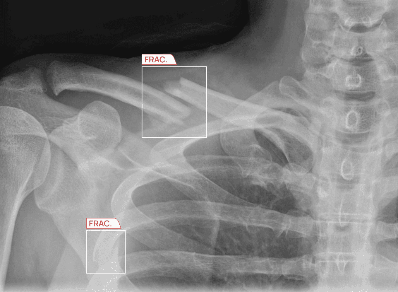
A hip fracture is considered one of the most dangerous fractures to miss because delayed diagnosis and treatment can lead to serious complications, particularly in older adults.
A missed diagnosis can turn a treatable injury into a serious complication that may impair mobility, independence, and quality of life.
Despite their severity, between 2% and 10% of nondisplaced femoral neck fractures may not be visible on initial radiographs.¹
In this article, we examine why that is, the consequences of delayed hip fracture diagnosis, and how AI hip fracture detection tools can help hospitals identify these cases.
Key takeaways
- Between 2% and 10% of nondisplaced femoral neck fractures may not be visible on initial X-rays.¹
- Delayed diagnosis can increase complications, surgical delays, and mortality risk in older patients.
- AI for hip fracture detection in the emergency department can help radiologists identify subtle fracture signs.
- AZtrauma supports fracture detection by identifying fractures, dislocations, and joint effusions on radiographs across the musculoskeletal system.
How hip fractures show up as nothing on the X-ray
Consider a scenario that occurs frequently at a healthcare center and among older patients.
A 70-year-old man arrives at the emergency department reporting hip pain and a restricted range of motion after suffering from a fall.
The radiologist on-site interprets an X-ray, but the radiograph shows no acute findings, so the man is discharged home with a diagnosis of a hip contusion.
But just 4 days later, he returns by ambulance as the condition has deteriorated. That is when he undergoes an MRI, and the clinicians identify a nondisplaced femoral neck fracture that was present all along.
The delay in diagnosis and treatment significantly complicates the clinical course.
This scenario illustrates a recognized diagnostic challenge. Between 2% and 10% of nondisplaced femoral neck fractures may not be visible on initial plain radiographs.¹ A Dutch teaching hospital analysis found that the hip and pelvis accounted for 37.3% of missed fractures in older emergency department patients, the highest proportion of any anatomical region in the study.² Missed fractures and delays in surgery are important concerns in older patients because of their association with adverse outcomes.
Hip fracture and fall statistics among older adults
There are multiple reports that highlight the scale of hip fractures in older adults and their consequences. It is a major public health concern, and here is why:
- According to the Centers for Disease Control and Prevention, nearly 319,000 older adults are hospitalized for hip fractures annually in the United States, with 88% of these fractures caused by falls.³
- Between 1999 and 2023, a study using CDC WONDER reported 334,905 deaths in US adults aged 65 and older were attributed to hip fractures. The study also highlighted an age-adjusted mortality rate of 23.91 per 100,000 in 2023 alone, with the highest rates concentrated in nonmetropolitan areas, the Midwest, and adults aged 85 and above.⁴
- Data from the University of Maryland Medical Center highlight the hip fracture mortality rates in older adults, which are 7% after 30 days, 15% after 90 days, and a significant 30% after 1 year.⁵
Moreover, a study of US mortality data identified 323,534 deaths associated with femoral neck fractures between 1999 and 2023.⁶ Let us learn more about why these fractures can be missed.
Why are hip fractures disproportionately missed in the emergency department?
There are three converging factors that create a diagnostic gap in hip fracture detection on plain X-ray, and they interact in ways that amplify each other's effects.
- Imaging limitation: Between 2% and 10% of nondisplaced femoral neck fractures may not be visible on initial plain radiographs.¹ MRI is the reference standard for detecting occult hip injury, but it is not the first-line modality in a high-volume emergency department. Patients presenting with hip pain after a fall are typically imaged with radiography, and a proportion of those radiographs will appear normal even when a fracture is present.
- Disproportionate miss rate in older patients: A Dutch teaching hospital analysis found that the hip and pelvis accounted for 37.3% of all missed fractures in older ED patients, the highest proportion of any anatomical region in the study.²
- Timing of missed diagnoses: The same analysis found that 49% of all missed fractures across age groups occurred between 4 PM and 9 PM.² This finding highlights the importance of maintaining reliable diagnostic processes throughout the day and evening. We discussed this in detail in our guide Why Emergency Fractures Are Most Likely to Be Missed at Night.
Why nondisplaced femoral neck fractures can be missed
Nondisplaced femoral neck fractures can be difficult to identify on initial radiographs, making early diagnosis particularly important.
Between 2% and 10% of nondisplaced femoral neck fractures may not be visible on plain radiographs at initial presentation.¹
In older adults, a missed fracture can delay treatment and contribute to adverse clinical outcomes.
In the case series, patients with initially nondisplaced femoral neck fractures verified on MRI had a 1-month mortality of 7%, a 1-year mortality of 20%, and a 2-year mortality of 31%.¹
How surgical delay affects hip fracture mortality
Once a hip fracture is detected, the American Academy of Orthopaedic Surgeons recommends performing surgery within 24 to 48 hours.⁷
That is because timely surgery can significantly influence the patient’s quality of life and mortality. Here are some studies that examine the relationship between surgical timing and mortality among patients with hip fractures.
- A meta-analysis of more than 190,000 patients found that surgery within 48 hours was associated with significantly lower mortality rates and fewer pressure ulcers than later surgical intervention.⁸
- A large prospective study collected data on patients between January 1989 and August 2021 and categorized time to surgery as less than 12 hours, 12–24 hours, 24–72 hours, or more than 72 hours. It found a significant increase in 30-day mortality when surgery was delayed beyond 12 hours after admission.⁹
- A 2026 study of 6,170 patients aged 60 and older found that patients whose transfer from the emergency department to a specialist orthopedic ward took more than 4 hours had 44% higher odds of 30-day mortality than those transferred within 4 hours.¹⁰
These studies highlight the importance of timely care for older adults with hip fractures. A missed diagnosis on the initial X-ray can delay further imaging and confirmation of the fracture, which may in turn delay surgical intervention.
The best way for emergency departments to mitigate this issue is to reduce the misdiagnoses in the first place. Here is how AI can help you do that.
The evidence base for AI hip fracture detection
AI tools for hip fracture detection are being evaluated for their ability to support fracture identification and radiograph interpretation. The following studies report their performance.
- The JAMA Network Open systematic review synthesized 39 studies covering 39,598 plain radiographs and found that the mean AI sensitivity for hip fracture detection reached 89.3%, and specificity was 87.5% across the studies. The authors of the study assessed these numbers as comparable to those of expert clinicians.¹¹
- Similarly, another meta-analysis in PLOS Digital Health, conducted across 66 studies and multiple fracture types, highlighted that hip fractures showed a pooled AI sensitivity of 92%, with a specificity of 90%.¹²
But how exactly do AI radiology tools help achieve these results? Let us find out.
How AI handles the fractures that plain X-ray misses
Deep-learning algorithms can be trained to identify imaging patterns associated with nondisplaced and occult hip fractures.
Convolutional neural networks (CNNs) can be trained on radiograph datasets to recognize imaging features associated with fractures, including subtle cortical disruptions and changes in trabecular bone patterns. The features detected depend on the system and its training data.
These subtle findings may be difficult for clinicians to identify, particularly when a fracture line is not clearly visible. AI systems may help flag findings for further review, but performance varies by system.
A 2025 study evaluating a deep-learning system specifically for nondisplaced femoral neck fractures on anteroposterior pelvic and hip radiographs reported an AUC of 0.991 and an overall accuracy of 94.8%. The system outperformed emergency physicians on the same image set.¹³
The study provides evidence of the evaluated system's performance on nondisplaced femoral neck fractures. Its findings should not be assumed to apply to other AI systems or to all real-world emergency department settings.
Joint effusion detection is a related imaging capability.
In some cases, an effusion may be considered alongside other clinical and imaging findings when assessing a patient for possible underlying injury. It does not independently establish the presence of an occult fracture.
The ability of an AI system to identify direct fracture findings or associated soft-tissue changes should be established through validation on the relevant hip and pelvis imaging datasets.
Why does the out-of-hours window matter specifically for hip fractures?
The timing of missed fractures is relevant when considering emergency department diagnostic processes across different shifts.
Mattijssen-Horstink et al. (2020) found that 49% of all missed fractures across age groups occurred between 4 PM and 9 PM.²
The study does not establish that the most diagnostically challenging fractures arrive during this period or that senior radiologist coverage is reduced at these times.
The finding nevertheless highlights the importance of maintaining reliable fracture-detection processes during evening hours.
AI systems do not experience human fatigue, although their diagnostic performance still needs to be validated in the intended clinical setting.
Consistent performance across operating hours should be assessed rather than assumed. For departments managing hip fracture presentations across extended shifts, validation of AI performance in out-of-hours settings is therefore important.
What to check when evaluating AI for hip and pelvis fracture detection
If you are considering a hip fracture detection AI tool for your hospital, here are a few criteria to keep in mind when making a choice.
Is the tool validated on elderly patient populations with osteoporotic bone?
Older patients with osteoporosis may have cortical thinning and trabecular changes that can make fracture interpretation more challenging. An AI tool's performance in this population should therefore be assessed using relevant validation data.
Does it cover all hip fracture types: femoral neck, intertrochanteric, subtrochanteric?
AI algorithms validated only on displaced fractures or on a single fracture pattern may not perform as well on nondisplaced femoral neck or intertrochanteric fractures on X-rays.
Does it maintain performance on nondisplaced hip fracture detection?
This is an important performance question for an AI pelvis fracture detection tool. Because nondisplaced fractures can be difficult to identify on plain radiographs, performance on these cases should be assessed separately from performance on displaced fractures.
Is it integrated at the PACS level with no added workflow steps?
Finally, consider whether the tool offers hip fracture PACS AI integration and does not complicate radiologists' workflow further.
Also, make sure that you are reading independent studies about the tool, not just the ones conducted by the vendor itself. To learn more, read our detailed guide on how to choose the best AI for fracture detection.
AZtrauma for fracture detection across the full musculoskeletal system
AZtrauma is an AI radiology solution for detecting fractures, dislocations, joint effusions, and osteoarticular lesions on X-rays. It has been validated at scale and is part of the AZmed Rayvolve AI suite, which is being used across 2,500+ healthcare centers.
Here are a few published evaluations conducted on AZtrauma and the results it achieved:
- Cohen et al. (2026), published in Radiography, evaluated AZtrauma across 258,373 X-rays from 100 medical centers in 26 countries. Across the full dataset, the algorithm achieved an AUC of 98.3%, a sensitivity of 97.4%, and a specificity of 96.4%.¹⁴
- Fu et al. (2024), published in Academic Radiology, evaluated AZtrauma in a multi-reader study of 24 emergency physicians and radiologists. With AI support, average reader sensitivity increased from 86.5% to 95.5% and reading time fell by 27%, with the largest gains for emergency physicians and non-musculoskeletal radiologists.¹⁵
To find more studies on AZtrauma, visit our scientific evidence section. Or, get in touch with our team to see how AZtrauma can be deployed at your hospital to support fracture detection across the musculoskeletal system.
Frequently asked questions
Can AI detect hip fractures on X-rays?
Yes. Studies have shown that AI systems can help detect hip fractures, including subtle and nondisplaced fractures that may be difficult to see on plain X-rays. Performance varies by system and should be evaluated using relevant clinical data.
Why are hip fractures missed on X-rays?
Some hip fractures do not create a clearly visible fracture line on plain radiographs. Osteoporotic bone, nondisplaced fractures, and emergency department workload can also contribute to missed diagnoses in older adults.
What is an occult hip fracture?
An occult hip fracture is a fracture that is not clearly visible on the initial X-ray but is later confirmed through MRI or CT imaging.
What is the hip fracture X-ray accuracy of AI?
Several studies report strong performance for AI hip fracture detection systems. A large review published in JAMA Network Open found mean AI sensitivity reached 89.3% and specificity reached 87.5% across nearly 40,000 radiographs. Another multi-study analysis reported a pooled AI sensitivity of 92% and specificity of 90% for hip fractures, with performance comparable to experienced clinicians in some settings.
Can AI detect nondisplaced femoral neck fractures on X-rays?
A 2025 study reported that a deep-learning system could detect nondisplaced femoral neck fractures on anteroposterior pelvic and hip radiographs.¹³ This finding applies to the system evaluated in that study and does not establish AZtrauma-specific performance.
What are the risks of delayed hip fracture diagnosis in the elderly in the USA?
Delayed diagnosis can lead to surgical delays, reduced mobility, longer hospital stays, and increased mortality risk in older adults.
How quickly should hip fracture surgery be performed?
Many guidelines recommend surgery within 24 to 48 hours because earlier treatment is linked to lower complications, whereas a hip fracture surgery delay increases mortality.
Does AI hip fracture detection work well on elderly patients with osteoporotic bone?
AI performance depends on the training data. Performance in older patients and those with osteoporotic bone should be established through validation in these populations.
Which type of hip fracture is most commonly missed on X-rays?
Nondisplaced femoral neck fractures can be difficult to identify because the fracture line may be very subtle on plain radiographs.
Can a patient still walk with a hip fracture?
Yes. Some patients with nondisplaced or occult hip fractures may still be able to walk, which can delay diagnosis and treatment. Older adults who experience a fall and develop hip pain should seek prompt medical assessment, even if they can still walk.
References
- Sundkvist J, Sjöholm P, Pejic A, et al. Clinical outcomes of patients with Garden I and II femoral neck fractures as verified on MRI: a retrospective case series. BMC Musculoskeletal Disorders. 2022;23:144.
- Mattijssen-Horstink L, Langeraar JJ, Mauritz GJ, et al. Radiologic discrepancies in diagnosis of fractures in a Dutch teaching emergency department: a retrospective analysis. Scandinavian Journal of Trauma, Resuscitation and Emergency Medicine. 2020;28:38.
- Centers for Disease Control and Prevention. Facts about older adult falls. CDC. Updated 2026.
- Tayyab M, Tanveer M, Ahmad Z, et al. Trends in hip fracture-related mortality among older adults in the United States from 1999 to 2023: a CDC WONDER analysis. Cureus. 2025;17(8):e90305.
- University of Maryland Medical Center. Mortality in hip fracture patients: identifying the prognostic value of troponin. UMMC physician brief.
- Faheem MSB, Saeed MU, Zeeshan N, et al. Trends in femoral neck fracture-related mortality in the United States: a nationwide retrospective study from 1999 to 2023. Medicine. 2026;105(8):e47754.
- American Academy of Orthopaedic Surgeons. Management of hip fractures in older adults: evidence-based clinical practice guideline. AAOS. 2021.
- Moja L, Piatti A, Pecoraro V, et al. Timing matters in hip fracture surgery: patients operated within 48 hours have better outcomes. A meta-analysis and meta-regression of over 190,000 patients. PLOS ONE. 2012;7(10):e46175.
- Warren M, Bretherton C, Parker M. Delay to surgery beyond 12 hours is associated with increased hip fracture mortality. European Journal of Orthopaedic Surgery & Traumatology. 2024;34(6):2973–2980.
- Katsanevakis M, Farag M, Norrish AR. Rapid transfer to specialist orthopaedic ward reduces mortality in hip fracture patients, but several factors reduce the ability to achieve the < 4-hour target. European Journal of Trauma and Emergency Surgery. 2026;52(1):54.
- Lex JR, Di Michele J, Koucheki R, et al. Artificial intelligence for hip fracture detection and outcome prediction: a systematic review and meta-analysis. JAMA Network Open. 2023;6(3):e233391.
- Jung J, Dai J, Liu B, Wu Q. Artificial intelligence in fracture detection with different image modalities and data types: a systematic review and meta-analysis. PLOS Digital Health. 2024;3(1):e0000438.
- Wang L, Zhang C, Wang Y, et al. Development and validation of a deep learning system for the detection of nondisplaced femoral neck fractures. Bioengineering. 2025;12(5):466.
- Cohen E, Ouertani MS, Beaumel P, et al. Performance of a complete AI radiographic suite across 258,373 X-rays from 26 countries: a worldwide evaluation. Radiography. 2026;32(4):103361.
- Fu T, Viswanathan V, Attia A, et al. Assessing the potential of a deep learning tool to improve fracture detection by radiologists and emergency physicians on extremity radiographs. Academic Radiology. 2024;31(5):1989-1999.
Regulatory information
US - Medical device Class II according to the 510K clearances. Rayvolve: is a computer-assisted detection and diagnosis (CAD) software device to assist radiologists and emergency physicians in detecting fractures during the review of radiographs of the musculoskeletal system. Rayvolve is indicated for adult and pediatric population (≥ 2 years).Rayvolve PTX/PE: is a radiological computer-assisted triage and notification software that analyzes chest x-ray images of patients 18 years of age or older for the presence of pre-specified suspected critical findings (pleural effusion and/or pneumothorax). Rayvolve LN: is a computer-aided detection software device to assist radiologists to identify and mark regions in relation to suspected pulmonary nodules from 6 to 30mm size of patients of 18 years of age or olderEU - Rayvolve: Medical Device Class IIa in Europe (CE 2797) in compliance with the Medical Device Regulation (2017/745). Rayvolve is a computer-aided diagnosis tool, intended to help radiologists and emergency physicians to detect and localize abnormalities on standard X-rays.Caution: The data mentioned are sourced from internal documents, internal studies and literature reviews. This material with associated pictures is non-contractual. It is for distribution to Health Care Professionals only and should not be relied upon by any other persons. Testimonial reflects the opinion of Health Care Professionals, not the opinion of AZmed. Carefully read the instructions for use before use. Please refer to our Privacy policy on our website For more information, please contact contact@azmed.co.AZmed 10 rue d’Uzès, 75002 Paris - www.azmed.co - RCS Laval B 841 673 601© 2026 AZmed – All rights reserved. MM-26-52