AI in radiology in India and the X-ray reporting gap

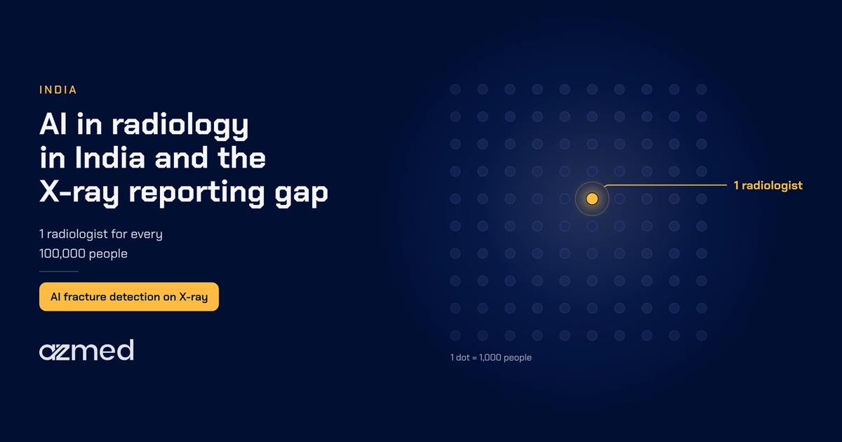
Every day, X-rays are taken at thousands of hospitals and diagnostic centers across India, and each one must be read by a radiologist, who then writes a report. With a limited number of radiologists, diagnostic chains, teleradiology hubs and hospital imaging departments face a practical question: how can they report more X-rays without lowering the quality of each report? X-rays can be taken in minutes, but reporting depends on how many radiologists are available. This reporting gap is what AI in radiology in India can help close.
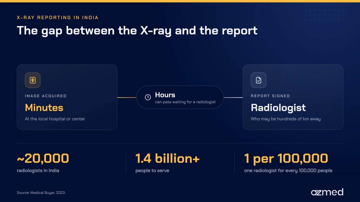
India has approximately 20,000 radiologists for more than 1.4 billion people, roughly one radiologist for every 100,000 people.¹ X-rays are low cost, fast to acquire and widely available. This is why X-ray reporting has become a key testing area for AI in India.
This article looks at where X-ray reporting in India is under strain and what AI can and cannot do on a radiograph. It also covers the evidence for AI fracture detection and offers cautions for buyers of AI radiology tools.
Key takeaways on AI in radiology in India
- AI's key promise is a shorter wait between taking an X-ray and receiving the report.
- Road traffic accidents and falls are frequent sources of musculoskeletal X-rays. This makes fracture detection one of the most pressing uses of AI on X-rays.
- AI works as a supplemental reviewer of radiographs. AI-enabled tools mark regions of interest on images, and the radiologist still makes all the decisions.
- In India, national guidance on medical device software, finalized in 2026, makes the regulatory status of AI radiology tools a key factor in purchasing decisions.
Pressure on X-ray reporting in India
The National Medical Commission (NMC) is expanding postgraduate training, with 3,224 MD (Radiodiagnosis) seats in its 2026-2027 seat matrix.² Yet each specialist needs 3 years of training after graduation, and imaging demand keeps increasing in the meantime. Thus, hiring alone cannot meet the radiology needs in India. This is one reason why AI has been included in the discussion.
Additionally, geography poses a significant challenge to the delivery of quality diagnostics in India.³ Local access to imaging may take only minutes. However, a patient can then wait hours for a report from a radiologist hundreds of kilometers away. AI support for radiology in India could bridge the gap and mitigate this situation.
In recent years, several public health programs have recognized the issue of delayed reporting and have acted to bridge this gap. Since 2015, the National Health Mission's Free Diagnostics Service Initiative has allowed states to provide teleradiology services and free X-rays. States can use in-house teams, public-private partnerships (PPPs) or a hybrid of both.⁴ These services expand access to imaging modalities. They have also concentrated large volumes of X-ray study requests on radiology teams in India, many of which are now looking at artificial intelligence (AI) technology.
Teleradiology carries a large share of the load
Teleradiology moves imaging studies from where they are taken to where a radiologist is located. It has become a main route for reporting X-ray studies in India. Many teleradiology companies now operate from central hubs. These hubs interpret images for multiple hospital systems and clinics across geographical areas, often at all hours. Therefore, teleradiology hubs are expected to be early adopters of AI technology in radiology in India.
Hubs face two main challenges. First, as demand for X-ray studies grows, so does the queue, because volume rises faster than staff can accommodate. Second, the hub's radiologists read numerous studies during their shift. These studies come from different imaging equipment, with varying image quality and from various patient demographics. Hubs therefore need consistent interpretations from all their radiologists. AI radiology tools for teleradiology in India can directly support consistent results. They can also help studies move through the queue faster.
Where trauma X-rays come from in India
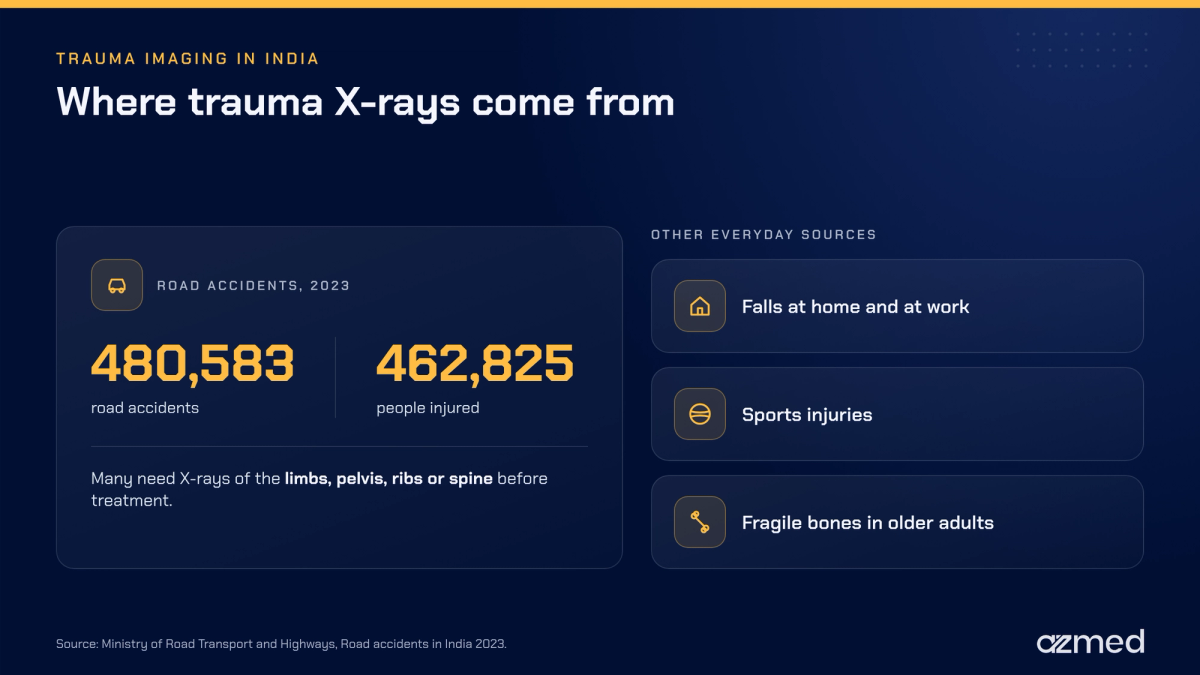
A significant share of urgent X-ray requests in India are for trauma. The Ministry of Road Transport and Highways (MoRTH) reported 480,583 road accidents in India in 2023, resulting in 172,890 fatalities and 462,825 injuries.⁵ Many of these injured patients will require limb, pelvis, rib or spine X-rays before clinicians can establish a treatment plan. Therefore, trauma X-ray examinations are a priority use case for AI in radiology in India.
However, many injuries result from sources other than crashes. Injuries at home and at work, during sporting activities and from fragile bones in older adults all add to the volume in emergency departments and orthopedic clinics. Most injured patients will first receive a plain X-ray as part of their workup. As a result, trauma creates a substantial volume of X-ray examinations.
What makes fractures easy to miss
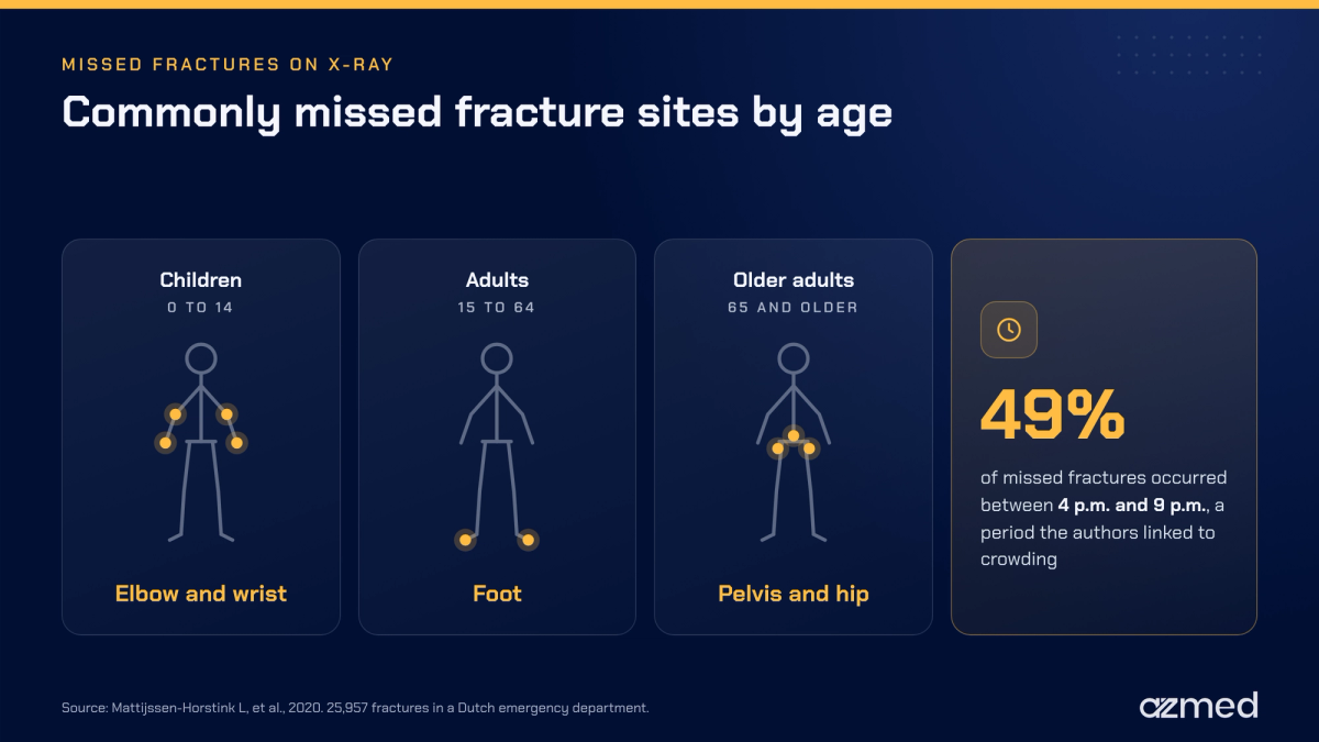
Fractures account for a significant portion of the findings on musculoskeletal X-ray examinations. Some of them are subtle in appearance, which makes them difficult to detect. A large study looked at 25,957 fractures treated in an emergency department in the Netherlands. Emergency physicians initially missed 1.1% of the fractures, and 9.3% of the missed fractures ultimately required surgery.⁶
Furthermore, the study showed that 49% of the missed fractures occurred between 4 p.m. and 9 p.m. Its authors linked this period to crowding in the department. The most commonly missed sites were the elbow and wrist in children, the foot in adults, and the pelvis and hip in individuals aged 65 and older.⁶ Although the study took place in the Netherlands, the same patterns of busy periods and subtle fracture appearance are likely to apply in high-volume settings in India.
For a diagnostic chain or teleradiology hub, a missed fracture leads to additional visits, delayed treatment planning and a loss of confidence from the referring physician. Avoiding these negative outcomes is the primary use case for AI in radiology in India on X-ray imaging.
What AI in radiology in India does on an X-ray
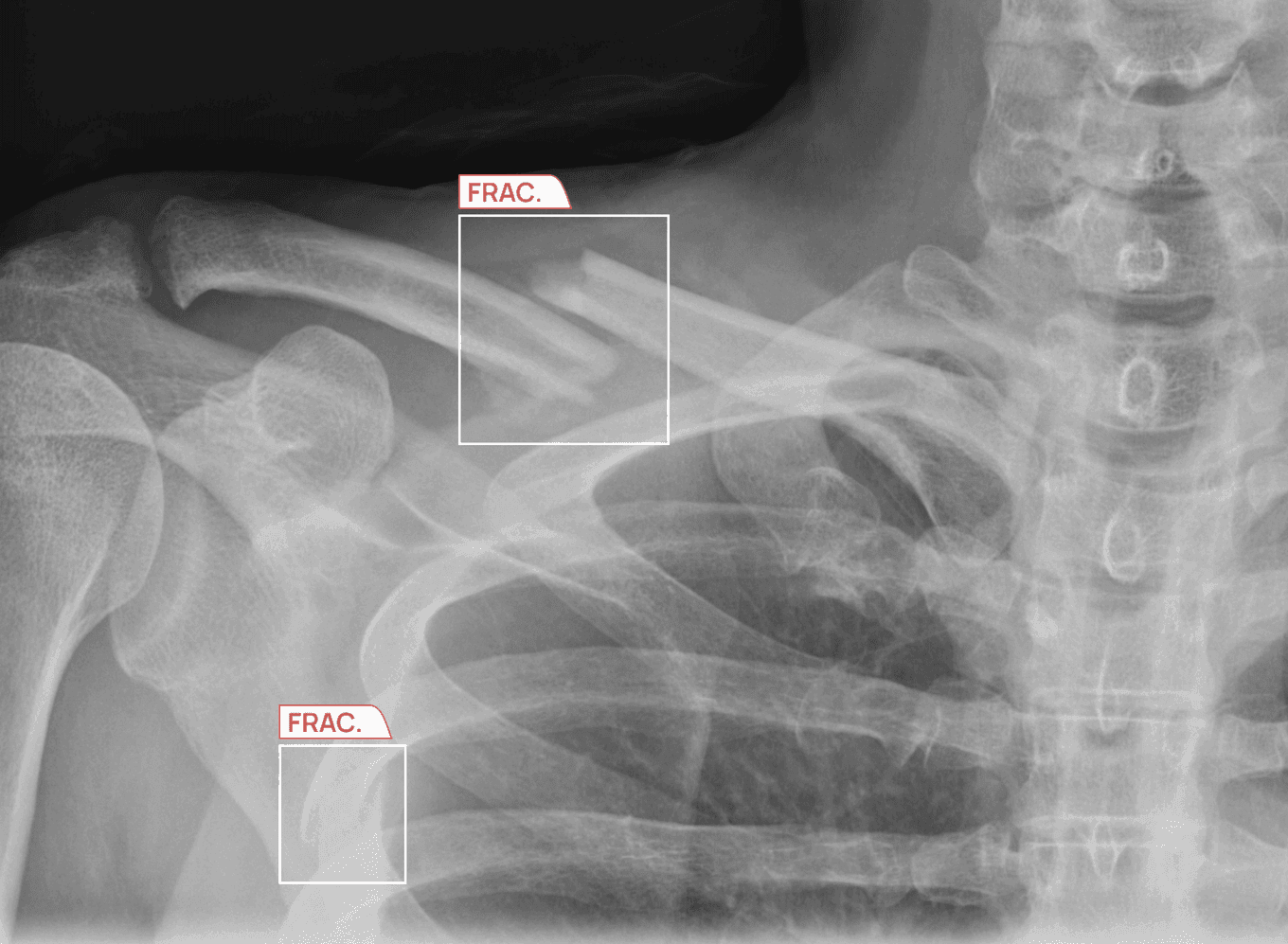
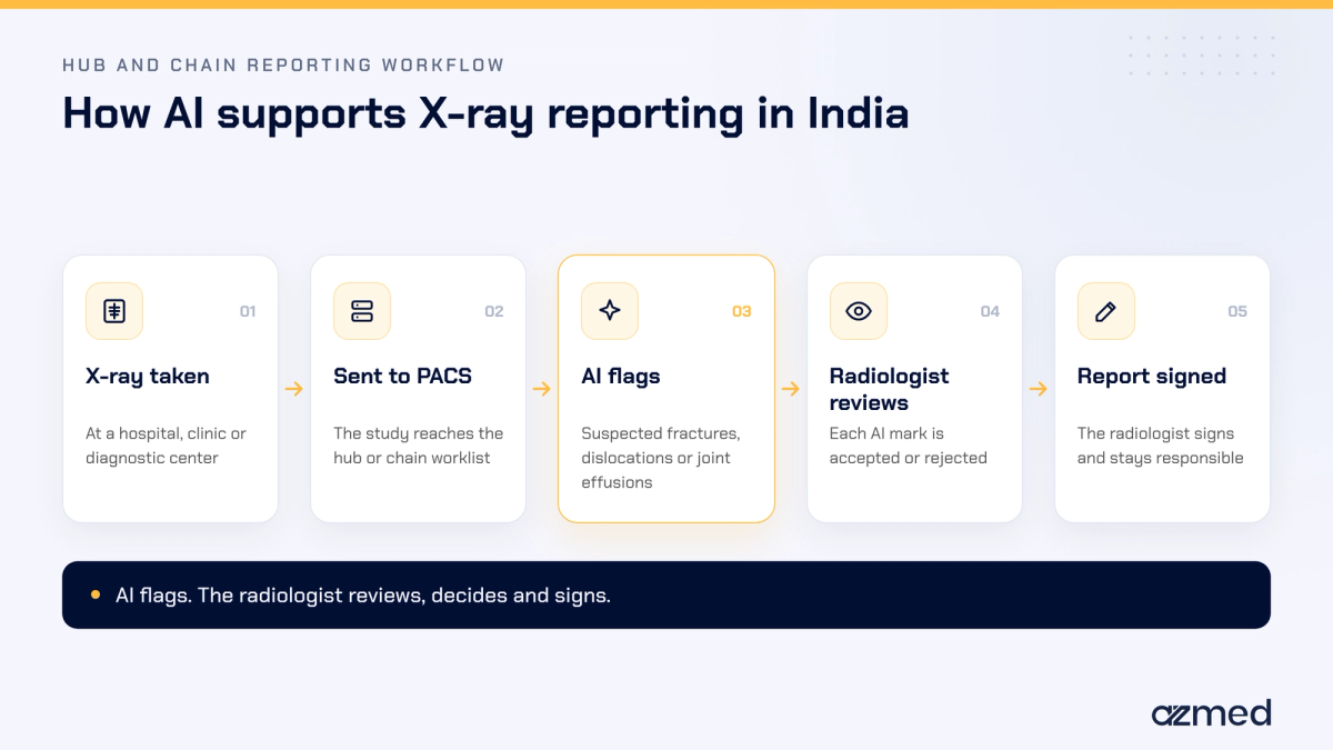
AI in radiology in India supports radiologists with analytic software that analyzes an X-ray image for potential abnormalities. The software highlights any area on the image where something could be wrong, e.g., a fractured bone or dislocated joint.
Next, radiologists review the marked regions on the X-ray image as they would any other part of the image. They then decide whether these findings are valid. After that, they create and sign a report on what they found during their review of the image.
In practice, AI can provide additional support to reporting teams in three areas.
- First, AI provides an additional layer of review for every X-ray study. During a long shift, the radiologist can quickly scan for marked areas of concern. They can check for subtle fractures in the wrist, elbow, hip or rib cage that may otherwise go unnoticed.
- Second, AI creates confidence in normal X-ray studies. Many X-rays in a trauma workflow are normal. When AI finds nothing suspicious in a study, the radiologist still reviews it, with the confidence that another check has taken place.
- Lastly, AI creates consistency across all facilities. Chains and hubs in India can give each radiologist the same level of support. This holds regardless of the X-ray machine, the time of day or the expertise of the radiologist reading the study.
It is equally important to understand what AI does not currently do in radiology in India. Radiologists still sign the reports, and treatment decisions remain with clinicians. They also keep both clinical and legal accountability for the reports they sign.
Where AI fits in an Indian reporting workflow
In India, reporting workflows in hubs and chains follow a similar path. Images flow from the X-ray machine to a picture archiving and communication system (PACS), and radiologists read from a centralized worklist. AI needs to fit into this workflow. A separate application or login would only add delays. Therefore, the AI tools for radiology that work best in India send their output directly into the PACS workflow.
What the evidence shows for AI fracture detection
Buyers interested in AI in radiology in India should consider two types of evidence. First, there is the accuracy of the AI tool when used on its own. Second, there is how the AI tool performs in actual radiological reporting.
Standalone accuracy across countries
AZmed's AZtrauma illustrates the first type of evidence. A retrospective review covered 258,373 radiographs from medical centers in 26 countries across five continents, collected between 2022 and 2025. AZtrauma achieved an area under the curve of 98.3%, with a 97.4% sensitivity and a 96.4% specificity for musculoskeletal findings.⁷ In this study, two readers read each image, and a third senior reader resolved any disagreements. AZtrauma showed robust performance across all subgroups of age, sex and country.⁷
The diversity of populations and equipment across India makes broad validation important for AI radiology tools. For example, image quality, technology and patient populations differ from one center to another. A radiology AI application validated in many countries and environments gives Indian buyers more confidence than one validated in a single country or population.
Real-world impact on reporting time
In addition to accuracy, reporting teams need to know how quickly urgent findings, such as a fracture, reach the referring physician. SimonMed Imaging is a large radiology group in the United States with multiple outpatient centers. It presented a pilot program at the RSNA 2023 Scientific Assembly and Annual Meeting, covering 50,682 X-ray examinations with AZmed's AI fracture detection. The mean turnaround time for fracture-positive examinations fell from 47.5 hours to 8.5 hours.⁸
These findings come from a U.S. study, and radiology practice in India is different. The AI results cannot simply be transferred to Indian radiology practice. Therefore, hospitals and groups that implement AI in radiology in India will need to assess the effects on their own volumes and operations.
Regulatory environment for AI in radiology in India
In 2026, the Central Drugs Standard Control Organisation (CDSCO) in India finalized guidance on registration and market access for medical device software. This followed a draft issued in 2025.⁹ The guidance places medical device software within the risk classes of India's Medical Device Rules, 2017. It also provides pathways for test licensing, clinical investigations, manufacture and import.⁹ The guidance is now the reference point for registering AI applications for radiology in India.
Under this framework, the manufacturer of AI-enabled radiology software for India has to explain its training, validation and testing datasets. It must also describe the demographic distribution and geographic origin of those datasets. In addition, the manufacturer must explain how it has considered bias, generalizability and robustness across subpopulations relevant to India.⁹
Consequently, this has a direct implication for the buyer. An AI radiology application may already have CE marking in Europe or FDA clearance in the United States. It may still need registration under CDSCO rules before routine clinical use in India. Therefore, anyone considering an AI product for radiology in India must ask each vendor two things. What is the product's status with CDSCO, and what data show its performance on Indian patients?
Questions for evaluating AI in radiology in India
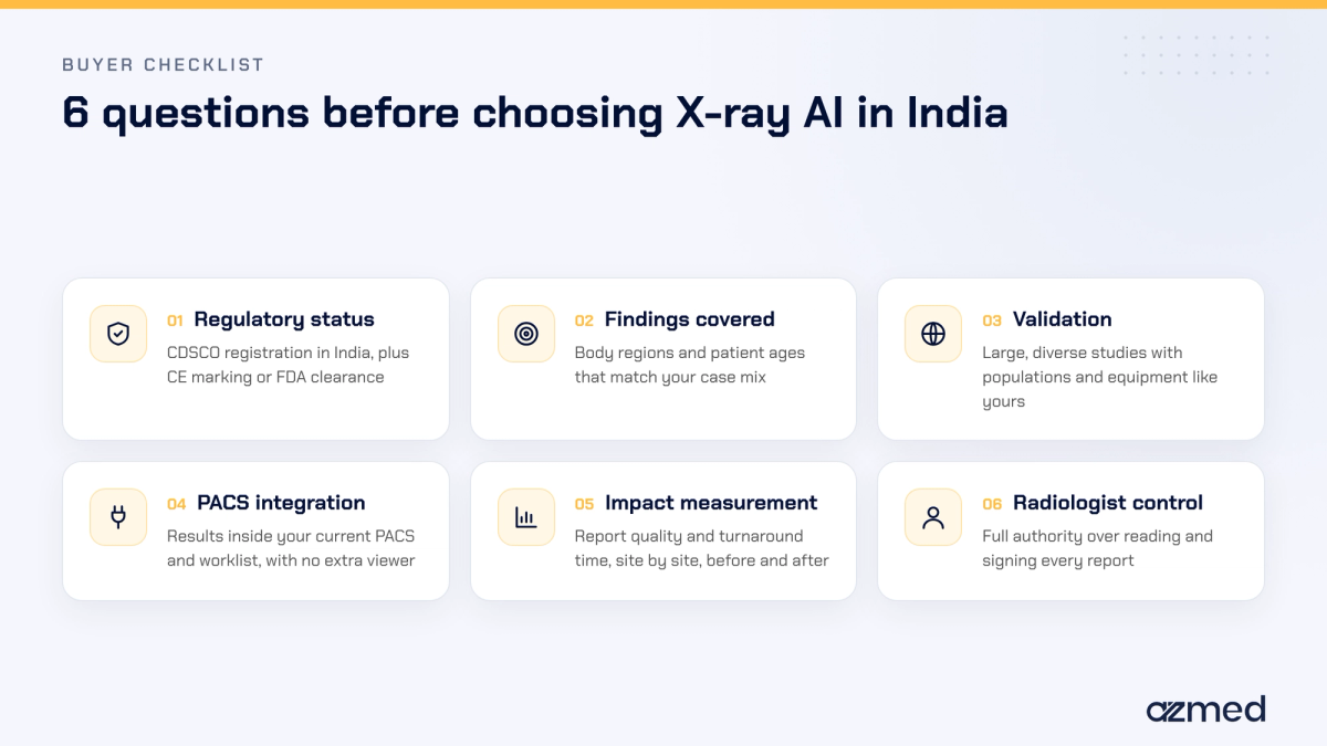
Diagnostic chains, teleradiology hubs and hospitals can use the following six questions to compare AI-based options in radiology in India.
- What regulatory status does the AI product hold for radiology use in India under CDSCO regulations, and what other certification (e.g., CE marking or FDA clearance) does it hold?
- Which findings, body areas or types of patients does the AI product cover, and are those similar to the patient types seen at our sites?
- How large and diverse were the validation studies, and do they have a population and equipment similar to ours?
- Does the AI product integrate with our current PACS and worklist, or does it need a separate viewer?
- How do we measure the impact of AI on quality and turnaround time (TAT) of reports at each site before and after deployment?
- Do radiologists retain full authority for reviewing and signing the reports?
The answers help separate AI products that fit radiology practice in India from those that only excel in a demo setting.
An example of AI in radiology in India at scale
Large radiology providers in India have started to test AI in their reporting workflows. In October 2026, Krsnaa Diagnostics announced a strategic AI collaboration with AZmed. The collaboration will evaluate the performance of AZtrauma for detecting fractures, joint effusions and dislocations on X-ray images.¹⁰ Krsnaa has over 2,000 X-ray centers across 19 states and union territories, with about 5 million X-ray reports issued each year.¹⁰ It also has a teleradiology hub in Pune that is recognized by the American College of Radiology.¹⁰
“With approximately 5 million X-rays reported every year, Krsnaa has the scale and operational experience to evaluate how AI-assisted radiology can contribute to more efficient workflows. Our collaboration with AZmed allows us to explore the potential of globally developed technology within the Indian healthcare ecosystem, while maintaining the central role of our radiologists in clinical decision-making.”
Dr. Madhuri Ghate, Vice President - Radiology, Krsnaa Diagnostics Limited
The collaboration is subject to successful evaluation, regulatory approvals and implementation.¹⁰ Structured evaluation at this scale shows how AI in radiology in India is likely to grow.
How AZmed supports AI in radiology in India
Alongside its collaboration with Krsnaa, AZmed develops Rayvolve, its AI suite of tools for X-ray imaging. Rayvolve's AZtrauma application detects fractures of the upper extremity, lower extremity, pelvis, rib cage and spine. It also detects dislocations and joint effusions in selected joints. AZtrauma is CE-marked in Europe and FDA-cleared in the US. In addition, AZmed solutions are currently used in more than 2,500 healthcare facilities around the world.¹⁰
The radiology teams at Krsnaa Diagnostics in India are currently evaluating AZmed's AI, AZtrauma. Before the technology can be used in routine patient care in India, it must first be registered with CDSCO. AZmed's AI integrates with existing PACS and worklists and marks suspected findings. All final decisions stay with the reporting team. Do you work in a diagnostic chain, teleradiology hub or hospital imaging center? To learn how AI in radiology in India fits within your X-ray workflow, please contact our team.
Frequently asked questions
What are the top radiology AI companies in India?
At this time, no official ranking exists for radiology AI companies in India. Only two groups of companies are typically compared: Indian companies that develop AI technologies for X-ray, CT or MRI, and international companies with AI applications already holding CE marking or FDA clearance. In October 2026, the Paris-based company AZmed, whose Rayvolve AI software suite has received a total of six FDA clearances, began working in India through a strategic collaboration with Krsnaa Diagnostics. A good place to start your search is by looking for AI companies with CDSCO registration, published literature about their products and references from current customers.
Can AI be used in radiology?
Yes, both Europe and the US have classified AI software for use in radiology as a medical device and primarily authorize AI as a supplemental tool that highlights areas of concern for interpretation by clinicians. In the United Kingdom, NICE (the National Institute for Health and Care Excellence) has issued a recommendation for AI-based fracture detection in urgent care X-ray applications, pending further evidence collection. In India, the Medical Device Rules, 2017 require that AI applications intended for routine clinical use be registered with the CDSCO. Clinicians always keep the authority to make the final medical decisions based on the findings of AI products.
How is AI used in radiology?
In radiology, AI applications focus on specific, defined tasks on medical images (X-ray, CT, MRI). Common AI functions within radiology include fracture detection, detection of chest X-ray abnormalities, timely notification of urgent studies (e.g., pneumothorax), measurements, estimation of bone age in pediatric patients and CT lung nodule detection. In every case, radiologists make the final interpretation decisions after being provided with AI product suggestions.
Is AI actually going to replace radiologists?
There is no data currently available to suggest that AI will be replacing radiologists any time soon. Existing AI tools are limited to performing specific individual tasks (e.g., detecting a fracture) and do not incorporate a patient's complete clinical history, but the nature of the work performed is shifting. As machines take over many of the repetitive initial tasks, radiologists can devote more time to complex cases, consultations and procedures.
What are 7 types of AI?
There are seven different types of AI, which are generally categorized into two major classifications. Based on capability, AI is divided into narrow AI, general AI and superintelligent AI. Based on functionality, AI can be defined in one of four ways: reactive machines, limited memory, theory of mind and self-aware AI. At present, only narrow AI, reactive machines and limited memory systems exist, with all other types being merely theoretical. In the context of medical imaging, AI trained for specific tasks, such as detecting fractures on X-rays, is classified as narrow AI.
Are radiographers safe from AI?
The current status of AI's impact on the profession of radiography does not suggest that radiographers will become obsolete due to advances in AI. The use of AI in radiology is mostly intended to support the interpretation of images after acquisition, and this does not replace the need for radiographers to perform their role: positioning patients, setting technical parameters for exposure, ensuring protection from unnecessary radiation and assisting patients during imaging examinations. Radiographers who are able to use AI effectively in their daily practice are likely to be highly valued within AI-capable departments.
How to learn AI in radiology?
For new learners who want to understand how AI is applied in the field of radiology, a practical method starts with learning the fundamentals of how algorithms are trained and validated, including sensitivity, specificity and area under the curve (AUC). Then, read articles from peer-reviewed journals about AI in radiology, for example Radiology: Artificial Intelligence. After that, take educational courses through a professional radiology society or an accredited university. The last step is to take part in an evaluation of an AI product at your hospital or diagnostic center, which will allow you to see how the AI product performs on cases from your region.
How is AI used in radiology in India?
In India, AI in radiology is primarily being used where image volumes are high. This includes large private diagnostic chains, teleradiology businesses and screening programs. Example use cases for AI in radiology in India are chest X-ray screening for tuberculosis, the detection of fractures on trauma X-rays and support for teleradiology hubs that report for remote sites. Many projects are still being evaluated, and AI products need CDSCO registration to be used routinely in clinical practice.
Is AI radiology software regulated in India?
Yes. In India, AI radiology software that is used for medical purposes is classified as a medical device, and as such, the Medical Device Rules, 2017 regulate AI software, with oversight by the Central Drugs Standard Control Organisation (CDSCO). Medical devices are classified into four risk classes (A to D). Class D medical devices undergo more intensive scrutiny and review than lower-risk classes. Since 2026, CDSCO guidance has asked AI developers for additional information about their training data and bias. Receiving a CE mark or FDA clearance does not replace the requirement to register with CDSCO in India.
How does AI help with the radiologist shortage in India?
AI will not add any radiologists to the workforce in India; however, it can help each radiologist interpret a larger number of X-rays each day with consistent quality. By marking images that contain possible findings, AI provides a second look at images during night shifts and peak times, when staff are most fatigued. For teleradiology hubs that serve small towns, this may help them maintain a stable turnaround time as their volume of reports continues to increase.
What should diagnostic chains in India look for in X-ray AI?
When selecting AI technology for radiology in India, diagnostic chains should look for solutions that have been registered with the CDSCO or are on a path to registration, have been validated in extensive multi-country studies, and can work within the PACS currently used by the organization. The AI technology should also identify the types of findings that drive report volumes, such as fractures and chest findings, fit the age range of the patients served, and include a way to track missed findings and turnaround time at each site.
How many radiologists are there in India?
As of 2023, according to figures cited in Indian medical news, India has approximately 20,000 radiologists for a population in excess of 1.4 billion people. That is approximately 1 radiologist for every 100,000 people in the country. The training of radiologists in India is on an upswing. In its seat matrix for 2026-2027, the National Medical Commission (NMC) lists 3,224 MD Radiodiagnosis seats, each leading to three years of training to become a radiologist.
Can AI detect fractures on X-rays?
Yes, AI is currently being used to aid in the detection of fractures, and this has become one of the most established uses of AI on X-ray images. In the United States, the Food and Drug Administration has cleared software such as AZmed's AZtrauma, which assists in detecting fractures, dislocations and joint effusions on X-ray images of patients aged 2 years and older. In European countries, such tools carry a CE mark. The software identifies a suspected fracture within the image, and the physician can then confirm or reject that finding.
How accurate is AI fracture detection on X-rays?
The level of accuracy of AI fracture detection on X-ray images depends on the population being tested and the particular product being used. A multi-country study of over 250,000 X-ray images using AZmed's AZtrauma showed 96.4% specificity and 97.4% sensitivity. Reader studies reflect another side of performance, i.e., whether clinical readers perform better when using AI; in AZmed's reader study data, the readers' area under the curve (AUC) went from 0.84 without AI to 0.89 with AI. Therefore, it is advised that purchasers ask vendors for both types of evidence.
How does AI fit into teleradiology in India?
AI technology fits into teleradiology in India as teleradiology hubs read X-ray studies sent digitally from district hospitals, clinics and diagnostic centers via the internet. Many of these X-ray studies are sent from different states and at night. The AI acts as a middle step, evaluating each study as it comes into the hub and applying marks that can be viewed in the hub's existing viewer. Each hub keeps the same worklist of studies, giving the radiologists a consistent second look across every site they work for.
Does radiology AI work with an existing PACS?
Yes. Many radiology AI tools are able to integrate with an existing PACS using the DICOM standard. The images to be evaluated are automatically sent to the AI system, and the AI's results are returned as an additional series or overlay alongside the original study. As a result, there is no change for the radiologist in the way studies are opened or reported, which makes adoption easier.
What is the future of AI in radiology in India?
The future development of AI in radiology in India is likely to be concentrated first in areas with a large volume of X-ray studies, such as teleradiology hubs, diagnostic chains and public-private diagnostic programs. The CDSCO guidance of 2026 creates clearer paths to registration for medical device software. Large-scale evaluations, such as the collaboration between Krsnaa Diagnostics and AZmed announced in October 2026, will be crucial in showing how AI technologies perform with Indian populations and the workflows associated with them.
Which X-ray findings can AI detect?
AI can detect fractures, dislocations and joint effusions on musculoskeletal X-rays, while on chest X-rays, AI can identify findings such as pneumothorax, pleural effusion, consolidation, an enlarged heart shadow and lung nodules. Additionally, certain systems can measure angles and lengths or estimate bone age. The specific findings that a given product may identify depend on its regulatory clearance, so buyers should check with the vendors.
Who is responsible for the report when AI is used in radiology?
The radiologist who signs the report is responsible for it, whether or not AI was used. AI output is a suggestion, not a diagnosis. The radiologist reviews every marked finding, accepts or rejects it, and never signs a report based on the AI result alone.
How can a hospital in India measure the impact of radiology AI?
A hospital in India can measure the impact of radiology AI with a before-and-after comparison over a fixed period, such as three to six months. Useful measures are the time from image acquisition to signed report, the number of findings corrected after review, the studies each radiologist reports per shift and feedback from referring doctors. Comparing results site by site shows where AI helps most.
References
- Medical Buyer. India has alarming ratio of one radiologist per lakh people. 2023.
- Medical Dialogues. 3,224 MD radiodiagnosis seats as per NMC seat matrix 2026-27. 2026.
- Chandramohan A, et al. Teleradiology and technology innovations in radiology: status in India and its role in increasing access to primary health care. The Lancet Regional Health Southeast Asia. 2023.
- National Health Systems Resource Centre. RFP for third-party evaluation of the Free Diagnostics Service Initiative. 2025.
- Ministry of Road Transport and Highways. Road accidents in India 2023. 2025.
- Mattijssen-Horstink L, et al. Radiologic discrepancies in diagnosis of fractures in a Dutch teaching emergency department: a retrospective analysis. Scandinavian Journal of Trauma, Resuscitation and Emergency Medicine. 2020.
- Cohen et al. Performance of a complete AI radiographic suite across 258,373 X-rays from 26 countries. Radiography. 2026.
- Raj S, et al. SimonMed Imaging pilot study presented at RSNA 2023. Reported in AuntMinnie, AI cuts time for radiologists reporting fractures on x-rays. 2023.
- Emergo by UL. India CDSCO finalizes guidance on medical device software. 2026.
- Krsnaa Diagnostics. Krsnaa Diagnostics announces strategic AI collaboration with global radiology AI innovator AZmed. 2026.
Regulatory information
US - Medical device Class II according to the 510K clearances. Rayvolve: is a computer-assisted detection and diagnosis (CAD) software device to assist radiologists and emergency physicians in detecting fractures during the review of radiographs of the musculoskeletal system. Rayvolve is indicated for adult and pediatric population (≥ 2 years).Rayvolve PTX/PE: is a radiological computer-assisted triage and notification software that analyzes chest x-ray images of patients 18 years of age or older for the presence of pre-specified suspected critical findings (pleural effusion and/or pneumothorax). Rayvolve LN: is a computer-aided detection software device to assist radiologists to identify and mark regions in relation to suspected pulmonary nodules from 6 to 30mm size of patients of 18 years of age or olderEU - Rayvolve: Medical Device Class IIa in Europe (CE 2797) in compliance with the Medical Device Regulation (2017/745). Rayvolve is a computer-aided diagnosis tool, intended to help radiologists and emergency physicians to detect and localize abnormalities on standard X-rays.Caution: The data mentioned are sourced from internal documents, internal studies and literature reviews. This material with associated pictures is non-contractual. It is for distribution to Health Care Professionals only and should not be relied upon by any other persons. Testimonial reflects the opinion of Health Care Professionals, not the opinion of AZmed. Carefully read the instructions for use before use. Please refer to our Privacy policy on our website For more information, please contact contact@azmed.co.AZmed 10 rue d’Uzès, 75002 Paris - www.azmed.co - RCS Laval B 841 673 601© 2026 AZmed – All rights reserved. MM-26-44