RSNA 2026 shows why radiologists must stay in control of AI

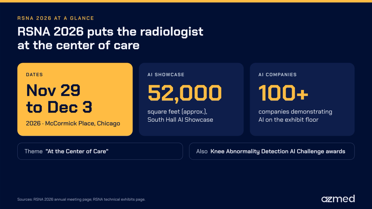
RSNA 2026 is going to take place from November 29 to December 3, 2026, at McCormick Place, located in Chicago. The theme for this year’s meeting is “At the Center of Care.”¹ The exhibit will include one of the largest displays of AI in radiology anywhere.² However, the theme of this year’s meeting puts the radiologist, not AI, at the center.
This tension is why we focus on the radiologists who will be walking through the exhibit floor. The question that radiologists must be asking themselves is not which tool seems to have the best technology. Instead, the main concern for them should be to stay in control by asking relevant questions about the technology.
Brief information on RSNA 2026
- RSNA 2026 is going to happen from November 29 to December 3, 2026.
- The location for RSNA 2026 is going to be McCormick Place, Chicago.
- The theme chosen for this year’s conference is “At the Center of Care.”¹
- An AI Showcase of approximately 52,000 square feet will anchor the RSNA 2026 exhibit, where more than 100 companies will demonstrate AI.²
- RSNA will present the RSNA Knee Abnormality Detection AI Challenge awards at the conference.
Reasons why US radiology needs AI to reduce workload
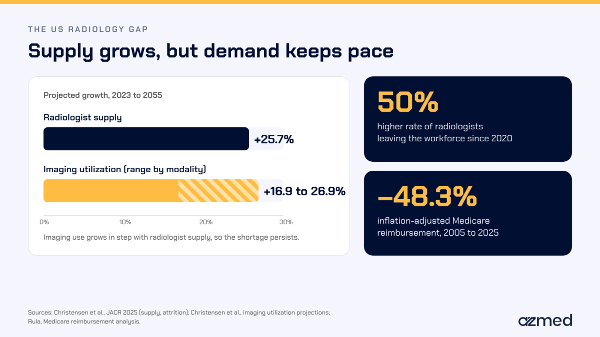
The radiologist shortage is a factor that no one can simply ignore in the US. Researchers project the supply of radiologists in the US to increase by 25.7% from 2023 to 2055.³ However, over the same period, the utilization of imaging may see an increase ranging from 16.9% to 26.9%, depending on the modality of imaging.⁴ Because supply and demand are in sync, the shortage will persist.
In addition, there are two more causes that make the shortage more severe. Radiologists have been leaving the workforce at a 50% higher rate since 2020.³ The amount Medicare reimburses radiologists, adjusted for inflation, also declined by 48.3% from 2005 to 2025. Meanwhile, the radiology workload continued to rise.⁵
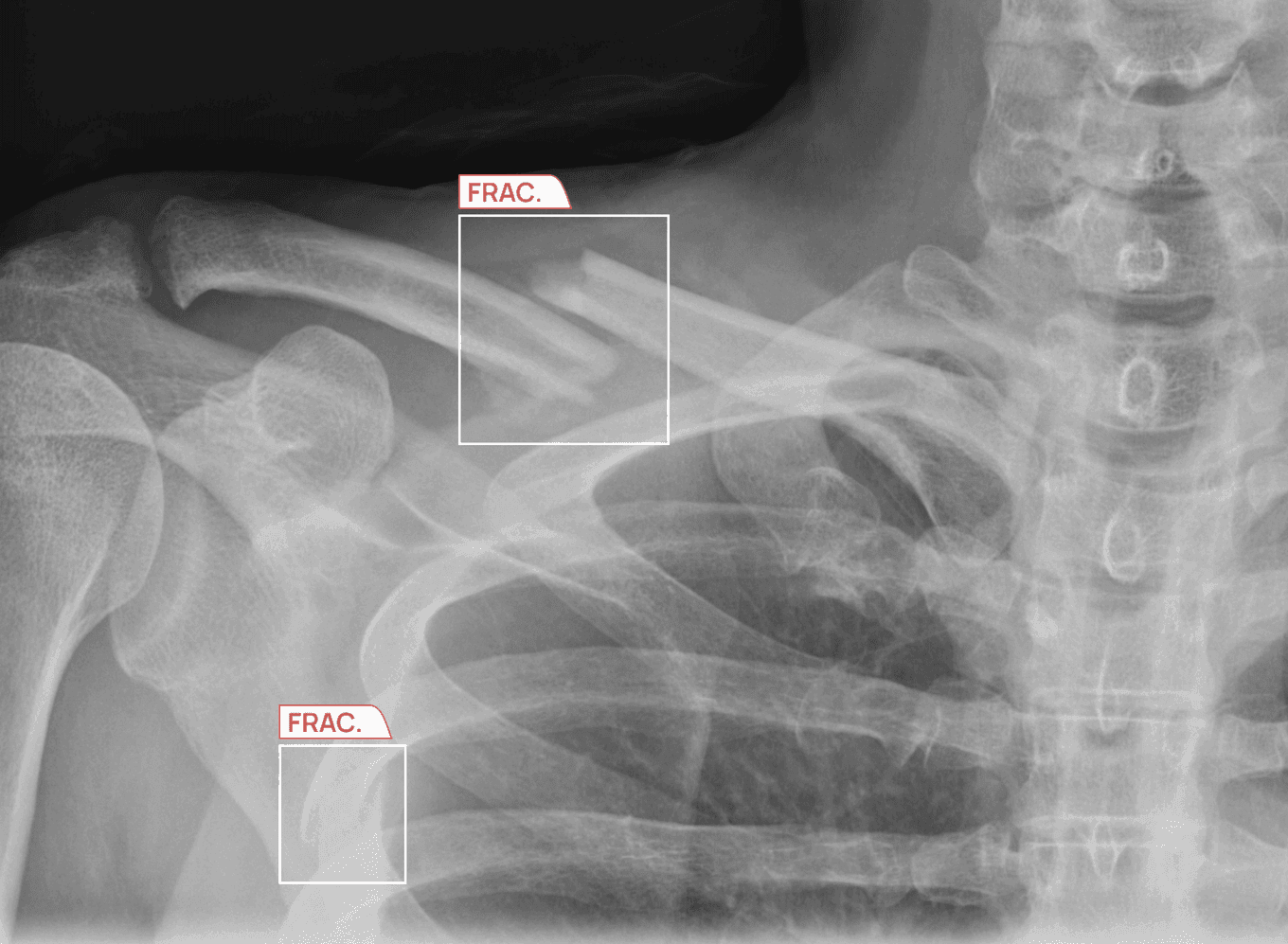
In practice, this shortage manifests itself as increased turnaround times and decreased time spent on each case. A fatigued reader dealing with a long worklist of cases at night is exactly where radiologists need additional support the most.
In conversations that we have had this year with radiology departments in the US, the same challenges came up. For instance, departments reported experiencing high turnover rates of radiologists and delays in recruitment. This led to the establishment of teleradiology services because staff were unable to keep up. Even with these backup measures in place, departments continue to seek out means to increase the productivity of radiologists. They are no longer questioning the need for assistance.
Where AI in radiology may fail to meet its purpose
Not all applications of AI technology provide benefits. For instance, a study conducted by MIT and Harvard of 140 radiologists and 15 chest X-ray tasks found that AI had differing effects on different readers. It also found that inaccurate AI predictions rendered radiologists less accurate. Moreover, knowledge and familiarity did not guarantee benefit from the technology for radiologists.⁶
So, while accuracy serves as the price of entry, it is not the only factor. Every additional interface, login procedure or unranked alert takes time. Multiply this time by thousands of studies, and the amount of time lost often proves to be massive.
Failing to solve the noise problem
An efficient application of technology works silently on negative studies and assertively when positive cases arise. Conversely, every false positive that radiologists need to dismiss takes attention away from the reader. It distracts them countless times each work shift.
Some US hospitals are already replacing the first AI tool they bought with a better one, instead of adding more tools on top.
How jurors determine liability regarding AI use
According to a study conducted in the US, researchers presented jurors in a simulated environment with a scenario where the AI indicated a brain hemorrhage. However, the radiologist was unable to detect it. Approximately three-fourths of jurors (72.9%) believed that the radiologist had committed malpractice. When researchers informed jurors about the AI’s error rate, the number of jurors who believed that the radiologist had committed malpractice decreased significantly.⁷
A follow-up study from 2026 investigated whether presenting the CT images to the radiologist before or after seeing the AI’s output impacted the jurors’ decisions. In this scenario, about 75% of jurors determined that the radiologist was at fault when the radiologist read the CT only once, after the AI flag. When the radiologist read the image before and after the AI flag appeared, that number dropped to 53%. This was nearly equal to the results when no AI was present.⁸
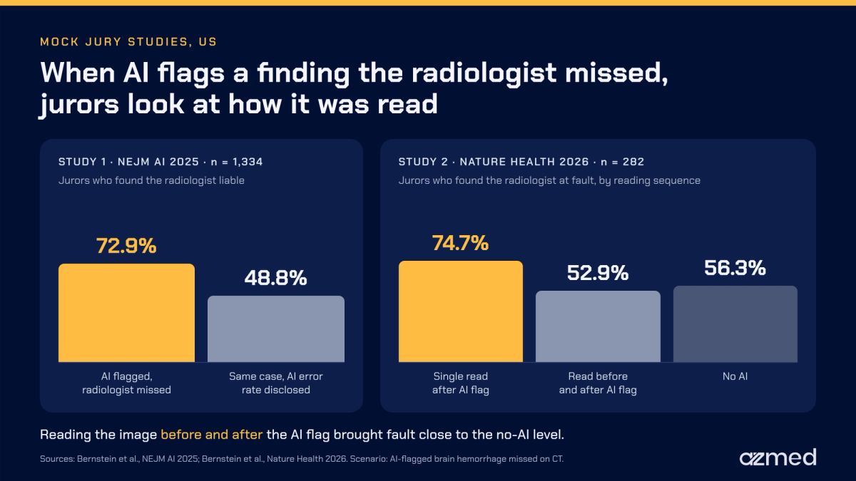
One likely reason for this difference is how the jurors interpret the reading sequence when the AI flag appears first as opposed to second. When the radiologist reads the image first and then checks the AI flag, it signifies an independent interpretation. However, the radiologist only checking the AI’s work might indicate that he or she checked the machine’s work rather than the patient’s image.
Malpractice risk management, not only IT planning, should now include where an AI system enters the sequence of reading for medical professionals. It should also include how teams document its error rates. Sometimes a radiologist overrides the output of an AI based on the knowledge or awareness that a limitation exists regarding the tool’s capabilities or reliability. In that case, there should be a clear record confirming this explicit act of the radiologist overriding the tool. Our exploration into this question included a more detailed analysis of the liability of radiologists who utilize AI.
How radiologists can keep the power of control over their use of AI
Control over the use of AI by a radiologist is not merely a phrase. Rather, it shows up in the design decisions a radiologist can check on the very first day of use. The following five design considerations are essential to control over an AI system:

AI to act as a second reader
A radiologist will read the study and unequivocally make the final decision on the study. AI will provide measurement, detection and suggestion as a secondary read of the study. It is not a substitute for the primary read.
The report is always editable and signed by the radiologist
Pre-populated reports save a radiologist typing. However, the system never signs them automatically at the completion of the review, and every word is editable. A report based on structured findings is much more efficient to edit and faster to complete.
Triage within the existing worklist
Critical findings should move up on the worklist of the radiologist, rather than appearing on a new screen for critical items. However, for triaging to run effectively, the AI must flag critical items with high specificity. Such flags must also be rare.
Connecting through existing systems
The ability to connect through the existing PACS system and standard HL7 messaging is essential for the use of AI in radiology. When the information technology department is able to complete a simple implementation, it gives control of the rollout to the radiologists. The sooner the radiologist is able to evaluate the AI tool based on actual studies, the better.
Using your own cases for validation
Each facility needs to validate how the software will work with its own equipment, patients and past studies prior to going live. In many cases, a brief test with retrospective data will answer most of the questions. A limited pilot at one site can then follow, prior to the larger rollout.
X-ray technology matters most
X-ray technology is where decisions surrounding AI use will matter most. It is one of the most frequently performed examinations within a diagnostic imaging department. When there is a small improvement in radiologist read time and confidence, it will translate into a large overall gain.
It also provides an opportunity to detect subtle abnormalities. Consider the identification of a subtle hairline fracture during a busy night shift. Or think of the identification of a small nodule during a routine chest X-ray examination. This is the reason why AI trauma detection, including AI fracture detection, and chest X-ray AI are the areas where many radiology groups start. They are also where groups continue to evaluate AI and test their control over it, study after study.
The future of RSNA 2026 AI
Visitors to the RSNA 2026 AI Showcase in Chicago can expect three transformations in artificial intelligence. These changes will move the technology closer to day-to-day clinical integration. At the same time, they will elevate the standards around control.

A single workflow for all studies
Many radiology practices no longer want to use one tool per finding. The new approach is to develop a single integrated workflow. It will encompass every step, from acquiring the image up until the draft report. Minimizing the number of handoffs of findings from one tool to another reduces the likelihood of losing clinical information along the way.
AI has now been able to expand to locations farther from a reading room, such as an urgent care center. The AI can mark suspected findings that support the clinician’s decision on what should happen with the patient. Meanwhile, the formal read occurs later. These flags can lead to the provision of a splint, a referral to a specialist, or the transfer of the patient to an emergency department.
The era of buying AI on promises is coming to an end. Purchasers of these services are no longer satisfied with promises of excellent performance. Instead, they are demanding to see a published and audited track record from AI vendors to determine their level of quality.
Federal and accredited organizations increasingly expect the same level of accountability from AI as from the complete reading process. Therefore, reports of performance by finding and patient subgroup, as well as surveillance of results after go-live, apply equally to AI.
If you are in the position of comparing RSNA AI companies, and AI X-ray companies in particular, these transitions offer an excellent checklist. In the end, the more AI does, the more the judgment of the radiologist will determine the care delivered.
AZmed will be exhibiting at RSNA 2026, Booth 5509
AZmed will be exhibiting the Rayvolve AI suite for X-ray at RSNA 2026, Booth 5509. Visitors will be able to see our suite in action in an actual reading environment. The demonstration will cover trauma and chest pathology detection, from triage to reporting. The radiologist maintains control at all stages of the process. Furthermore, AZmed representatives will be on hand to explain how the concept of control applies to your practice. We invite you to plan your visit to Booth 5509 or get in touch with our representatives.
Frequently asked questions
When and where is RSNA 2026?
November 29 to December 3, 2026, at McCormick Place in Chicago.
What is the RSNA 2026 theme?
The theme for RSNA 2026 is “At the Center of Care.” Radiology plays a major, central part in patient care.
How many AI companies will be at RSNA 2026?
More than 100 companies will demonstrate AI, many of them in the AI Showcase. The trends covered above list the AI technologies to look for.
Will AI technologies replace radiologists?
No. Utilization of imaging continues to increase as fast as the workforce grows. Currently, there is evidence to support the use of AI technology to augment radiologists.
Who will assume legal responsibility for a finding flagged by AI but overlooked by a radiologist?
Mock jury studies suggest that radiologists may bear liability for an AI-flagged finding that they missed. The reading sequence is an important factor, as explained in the liability section above.
References
- RSNA. Annual meeting announcement: rsna.org. 2026.
- RSNA. Technical exhibits announcement: rsna.org. 2026.
- Christensen EW, Parikh JR, Drake AR, Rubin EM, Rula EY. Projected US radiologist supply, 2025 to 2055. Journal of the American College of Radiology. 2025.
- Christensen EW, et al. Neiman HPI study of projected US imaging utilization to 2055. Journal of the American College of Radiology. 2025.
- Rula EY, et al. Neiman HPI editorial on AI and the radiology workforce. Journal of the American College of Radiology. 2026.
- Yu F, Moehring A, Banerjee O, Salz T, Agarwal N, Rajpurkar P. Heterogeneity and predictors of the effects of AI assistance on radiologists. Nature Medicine. 2024.
- Bernstein MH, Sheppard B, Bruno MA, Lay PS, Baird GL. Randomized study of the impact of AI on perceived legal liability for radiologists. NEJM AI. 2025.
- Bernstein MH, Sheppard B, Bruno MA, Lay PS, Baird GL. The radiologist-AI workflow and the risk of medical malpractice claims. Nature Health. 2026.
Regulatory information
US - Medical device Class II according to the 510K clearances. Rayvolve: is a computer-assisted detection and diagnosis (CAD) software device to assist radiologists and emergency physicians in detecting fractures during the review of radiographs of the musculoskeletal system. Rayvolve is indicated for adult and pediatric population (≥ 2 years).Rayvolve PTX/PE: is a radiological computer-assisted triage and notification software that analyzes chest x-ray images of patients 18 years of age or older for the presence of pre-specified suspected critical findings (pleural effusion and/or pneumothorax). Rayvolve LN: is a computer-aided detection software device to assist radiologists to identify and mark regions in relation to suspected pulmonary nodules from 6 to 30mm size of patients of 18 years of age or olderEU - Rayvolve: Medical Device Class IIa in Europe (CE 2797) in compliance with the Medical Device Regulation (2017/745). Rayvolve is a computer-aided diagnosis tool, intended to help radiologists and emergency physicians to detect and localize abnormalities on standard X-rays.Caution: The data mentioned are sourced from internal documents, internal studies and literature reviews. This material with associated pictures is non-contractual. It is for distribution to Health Care Professionals only and should not be relied upon by any other persons. Testimonial reflects the opinion of Health Care Professionals, not the opinion of AZmed. Carefully read the instructions for use before use. Please refer to our Privacy policy on our website For more information, please contact contact@azmed.co.AZmed 10 rue d’Uzès, 75002 Paris - www.azmed.co - RCS Laval B 841 673 601© 2026 AZmed – All rights reserved. MM-26-40